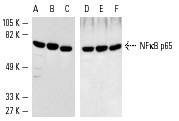
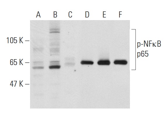
Western blot analysis of NFκB p65 in NIH/3T3 (A,D), A-431 (B,E) and K-562 (C,F) whole cell lysates. Antibodies tested include NFκB p65 (C-20): sc-372 (A-C) and NFκB p65 (C-20)-G: sc-372-G (D-F).
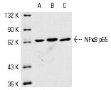
NFκB p65 (C-20)-G: sc-372-G. Western blot analysis of NFκB p65 expression in K-562 (A), A-431 (B) and NIH/3T3 (C) whole cell lysates.
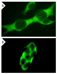
NFκB p65 (C-20): sc-372. (A) Immunofluorescence staining of methanol-fixed NIH/3T3 cells showing cytoplasmic staining. (B) NFκB p65 (C-20)-G: sc-372-G. Immunofluorescence staining of methanol-fixed A-431 cells showing cytoplasmic staining.
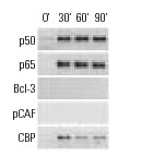
ChIP analysis of cofactor occupancy dynamics on the ICAM1 promoter in 293 cells in response to IL-1β treatment. Antibodies tested include NFκB p50 (C-19): sc-1190, NFκB p50 (E-10): sc-8414, NFκB p50 (H-119): sc-7178, NFκB p65 (C-20): sc-372, NFκB p65 (A): sc-109, NFκB p65 (H-286): sc-7151, Bcl-3 (C-14): sc-185, Bcl-3 (H-146): sc-13038, PCAF (C-16): sc-6300, PCAF (H-369): sc-8999, CBP (A-22): sc-369, CBP (C-1): sc-7300, CBP (C-20): sc-583, CBP (451): sc-1211. Data kindly provided by M.G. Rosenfeld and reproduced with permission from Baek et al., Cell 2002, 110: 55-67.
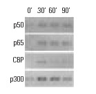
ChIP analysis of cofactor occupancy dynamics on the IL-8 promoter in 293 cells in response to IL-1β treatment. Antibodies tested include NFκB p50 (C-19): sc-1190, NFκB p50 (E-10): sc-8414, NFκB p50 (H-119): sc-7178, NFκB p65 (C-20): sc-372, NFκB p65 (A): sc-109, NFκB p65 (H-286): sc-7151, CBP (A-22): sc-369, CBP (C-1): sc-7300, CBP (C-20): sc-583, CBP (451): sc-1211, p300 (C-20): sc-sc-585, p300 (N-15): sc-584, p300 (H-272): sc-8981. Data kindly provided by M.G. Rosenfeld and reproduced with permission from Baek et al., Cell 2002, 110: 55-67.
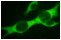
NFκB p65 (C-20): sc-372. Immunofluorescence staining of methanol-fixed NIH/3T3 cells showing cytoplasmic staining.
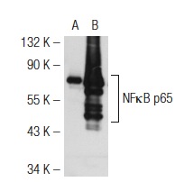
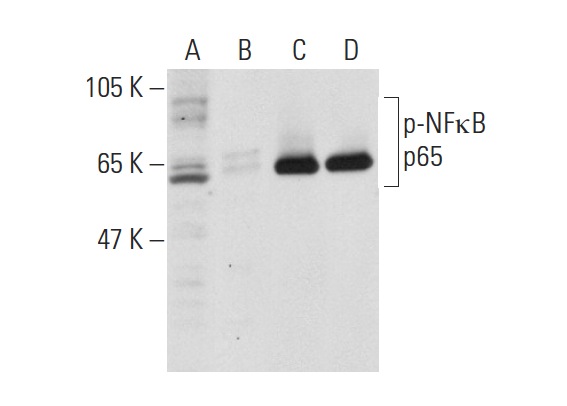
NFκB p65 (C-20): sc-372. Western blot analysis of NFκB p65 expression in non-transfected: sc-117752 (A) and mouse NFκB p65 transfected: sc-122027 (B) 293T whole cell lysates.
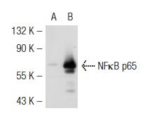
NFκB p65 (C-20)-G: sc-372-G. Western blot analysis of NFκB p65 expression in non-transfected: sc-117752 (A) and mouse NFκB p65 transfected: sc-122027 (B) 293T whole cell lysates.
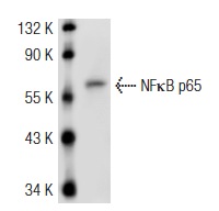
NFκB p65 (C-20)-G: sc-372-G. Western blot analysis of NFκB p65 expression in NIH/3T3 whole cell lysate.
Western blot analysis of NFκB p65 phosphorylation in untreated (A,D), TNFα and calyculin A treated (B,E) and TNFα, calyculin and lambda protein phosphatase treated (C,F) HeLa whole cell lysates. Antibodies tested include p-NFκB p65 (Ser 311): sc-33039 (A,B,C) and NFκB p65 (C-20): sc-372 (D,E,F).
Western blot analysis of NFκB p65 phosphorylation in untreated (A,C), and lambda protein phosphatase treated (B,D) K-562 whole cell lysates. Antibodies tested include p-NFκB p65 (Ser 311): sc-33039 (A,C) and NFκB p65 (C-20): sc-372 (B,D).
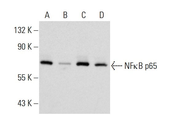
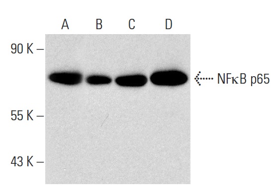
NFκB p65 (C-20)-G: sc-372-G. Western blot analysis of NFκB p65 expression in HeLa (A), MIA PaCa-2 (B), T24 (C) and SK-BR-3 (D) whole cell lysates.
NFκB p65 (C-20)-G: sc-372-G. Western blot analysis of NFκB p65 expression in non-transfected 293T: sc-117752 (A), mouse NFκB p65 transfected 293T: sc-122027 (B), Jurkat (C) and K-562 (D) whole cell lysates.
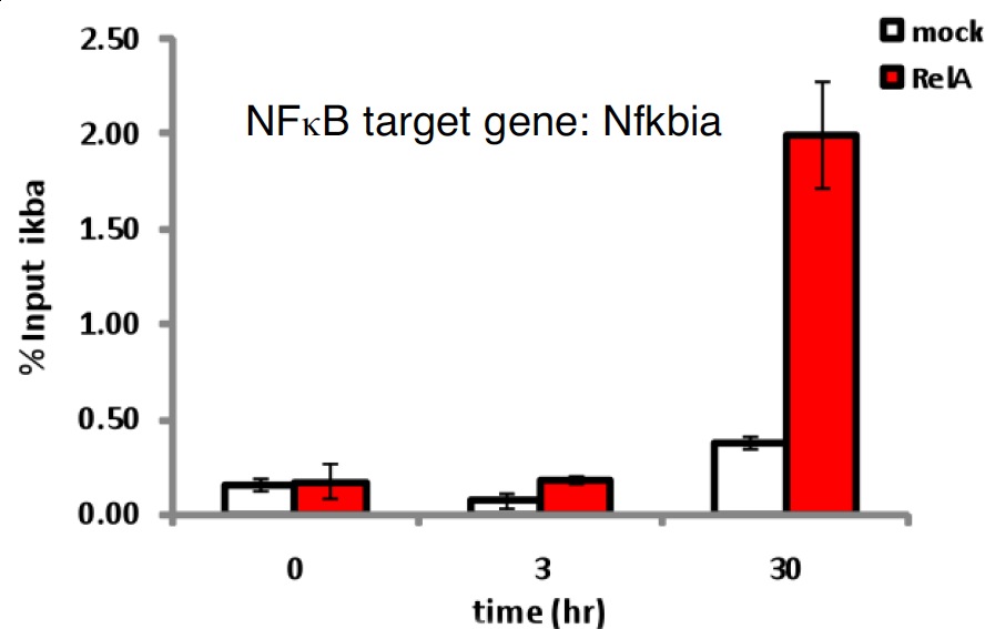
NFB p65 (C-20): sc-372. Quantitative RT-PCR data using primers spanning the promoter of the IkBa gene (Nfkbia, a known NFkB target) of ChIP samples prepared from murine splenic B-cells stimulated with 50ng/ml BAFF for indicated times. 5g of antibody and 107 cells were used for each sample. The specificity of the signal is confirmed by using mock IgG (sc-2025) and primers for an unrelated locus where NFkB does not bind (Not shown). Kindly provided by the Dr. Alexander Hoffmann Laboratory, University of California San Diego.