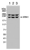
All lanes : Anti-Xrn1 antibody (ab70259) at 1/10000 dilutionLane 1 : Whole cell lysate from HeLa cells at 5 µgLane 2 : Whole cell lysate from HeLa cells at 15 µgLane 3 : Whole cell lysate from HeLa cells at 50 µgdeveloped using the ECL technique
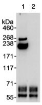
1 mg of whole cell lysate from HeLa cells was immunoprecipitated with ab70259 at 3ug/mg lysate (lane 1) or with normal rabbit IgG (lane 2). The subsequent blot was performed using ab70259 at 1ug/ml.
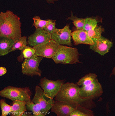
ICC/IF image of ab70259 stained DU145 cells. The cells were 4% formaldehyde fixed (10 min) and then incubated in 1%BSA / 10% normal goat serum / 0.3M glycine in 0.1% PBS-Tween for 1h to permeabilise the cells and block non-specific protein-protein interactions. The cells were then incubated with the antibody (ab70259, 5µg/ml) overnight at +4°C. The secondary antibody (green) was ab96899, DyLight® 488 goat anti-rabbit IgG (H+L) used at a 1/250 dilution for 1h. Alexa Fluor® 594 WGA was used to label plasma membranes (red) at a 1/200 dilution for 1h. DAPI was used to stain the cell nuclei (blue) at a concentration of 1.43µM.