Lane 1 : Anti-Wnt10b antibody (ab70816) at 2 µg/mlLane 2 : Anti-Wnt10b antibody (ab70816) at 4 µg/mlLane 1 : Human skeletal muscle tissue lysateLane 2 : Human skeletal muscle tissue lysateLysates/proteins at 15 µg per lane.
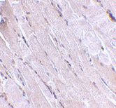
ab70816 at 2.5 µ/ml staining Wnt10 in human skeletal muscle tissue section by Immunohistochemistry (Formalin/ PFA fixed paraffin-embedded tissue sections).
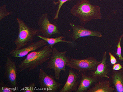
ICC/IF image of ab70816 stained HeLa cells. The cells were 4% formaldehyde fixed (10 min) and then incubated in 1%BSA / 10% normal goat serum / 0.3M glycine in 0.1% PBS-Tween for 1h to permeabilise the cells and block non-specific protein-protein interactions. The cells were then incubated with the antibody (ab70816, 5µg/ml) overnight at +4°C. The secondary antibody (green) was ab96899, DyLight® 488 goat anti-rabbit IgG (H+L) used at a 1/250 dilution for 1h.Alexa Fluor® 594 WGA was used to label plasma membranes (red) at a 1/200 dilution for 1h. DAPI was used to stain the cell nuclei (blue) at a concentration of 1.43µM.