Phospho-DAPP1/BAM32 (Tyr139) (D7G4G) Rabbit mAb
| Name | Phospho-DAPP1/BAM32 (Tyr139) (D7G4G) Rabbit mAb |
|---|---|
| Supplier | Cell Signaling Technology |
| Catalog | 13703 |
| Prices | $287.00 |
| Sizes | 100 µl (10 western blots) |
| Host | Rabbit |
| Clonality | Monoclonal |
| Isotype | IgG |
| Clone | D7G4G |
| Applications | WB IP |
| Species Reactivities | Human |
| Antigen | Monoclonal antibody is produced by immunizing animals with a synthetic phosphopeptide corresponding to residues surrounding Tyr139 of human DAPP1/BAM32 protein. |
| Description | Rabbit Monoclonal |
| Gene | DAPP1 |
| Supplier Page | Shop |
Product images
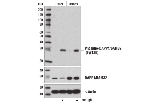 Western blot analysis of extracts from Daudi and Ramos cells, untreated (-) or treated with anti-IgM (12 μg/ml, 10 min; +), using Phospho-DAPP1/BAM32 (Tyr139) (D7G4G) Rabbit mAb (upper), DAPP1/BAM32 (D9K4O) Rabbit mAb #13598 (middle), and β-Actin (D6A8) Rabbit mAb #8457 (lower).
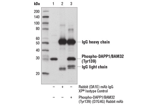 Immunoprecipitation of phospho-DAPP1/BAM32 (Tyr139) protein from Daudi cells treated with anti-IgM (12 μg/ml, 10 min) using Rabbit DA1E mAb XP ® Isotype Control #3900 (lane 2) or Phospho-DAPP1/BAM32 (Tyr139) Rabbit mAb (lane 3). Lane 1 is 10% input. Western blot analysis was performed using Phospho-DAPP1/BAM32 (Tyr139) (D7G4G) Rabbit mAb.