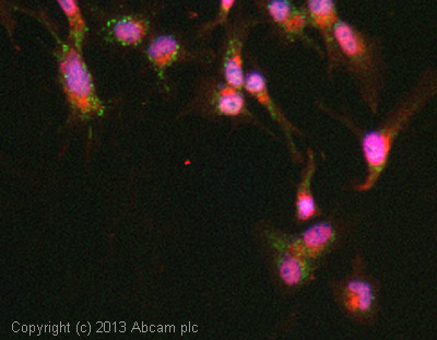
ICC/IF image of ab39250 stained HepG2 cells. The cells were 4% formaldehyde fixed (10 min) and then incubated in 1%BSA / 10% normal goat serum / 0.3M glycine in 0.1% PBS-Tween for 1h to permeabilise the cells and block non-specific protein-protein interactions. The cells were then incubated with the antibody (ab39250, 1µg/ml) overnight at +4°C. The secondary antibody (green) was ab96899, DyLight® 488 goat anti-rabbit IgG (H+L) used at a 1/250 dilution for 1h. Alexa Fluor® 594 WGA was used to label plasma membranes (red) at a 1/200 dilution for 1h. DAPI was used to stain the cell nuclei (blue) at a concentration of 1.43µM.
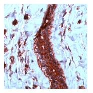
Human angiosarcoma stained with ab39250.
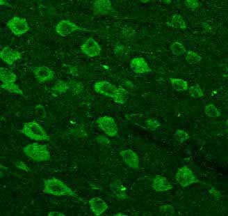
ab39250 staining VEGF in mouse brain tissue by Immunohistochemistry (Free floating sections). A heat mediated antigen retrieval step (100°C for 20 minutes) was performed using 10mM sodium citrate buffer, 0.05% Tween 20 pH 6.0. Samples were then incubated with the primary antibody at a 1/100 dilution for 72 hours at 4°C. An Alexa Fluor®488-conjugated goat anti-rabbit polyclonal was used as secondary antibody at a 1/500 dilution.ab39250 staining VEGF in mouse brain tissue by Immunohistochemistry (Free floating sections). A heat mediated antigen retrieval step (100°C for 20 minutes) was performed using 10mM sodium citrate buffer, 0.05% Tween 20 pH 6.0. Samples were then incubated with the primary antibody at a 1/100 dilution for 72 hours at 4°C. An Alexa Fluor®488-conjugated goat anti-rabbit polyclonal was used as secondary antibody at a 1/500 dilution.See Abreview