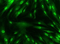
Immunocytochemistry image of ab110308-stained Human HDFn cells. The cells were paraformaldehyde fixed (4%, 20 min) and Triton X-100 permeabilized (0.1%, 15 min). The cells were incubated with ab110308 at 2 µg/ml) for 2 hours at room temperature or over night at 4°C. The secondary antibody was (green) Alexa Fluor® 488 goat anti-mouse IgG (H+L) used at a 1/1000 dilution for 1 hour. 10% Goat serum was used as the blocking agent for all blocking steps. Target protein locates mainly in nucleus and cytoplasm.
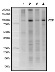
ab110308 pulls down the 89 kDa VCP protein in Human (lane 1), Rat (lane 2), and Mouse (lane 3) liver samples and Human HepG2 cultured cell lysate (lane 4). The identity of this protein was confirmed by mass spectrometry. This gel was stained with sypro ruby gel stain.
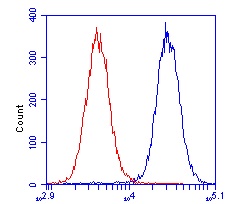
HL-60 cells were stained with 1 µg/mL ab110308 (blue) or an equal amount of an isotype control antibody (red) and analyzed by flow cytometry.