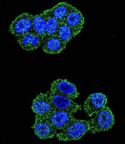
Confocal immunofluorescent analysis of Hela cells labeling UQCRFS1 with ab170470 at 1/10 dilution, followed by Alexa Fluor 488-conjugated goat anti-rabbit lgG (green). DAPI was used to stain the cell nuclei (blue).
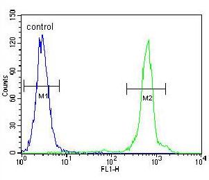
Flow cytometric analysis of Hela cells labeling UQCRFS1 with ab170470 at 1/10 dilution (right histogram), compared to negative control cells (left histogram). FITC-conjugated goat-anti-rabbit secondary antibodies were used for the analysis.
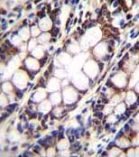
Immunohistochemical analysis of formalin-fixed, paraffin-embedded Human rectum tissue labeling UQCRFS1 with ab170470 at 1/10 dilution, followed by a peroxidase conjugated secondary antibody and DAB staining.
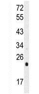
Anti-UQCRFS1 antibody - C-terminal (ab170470) at 1/100 dilution + HeLa cell lysate at 35 µg