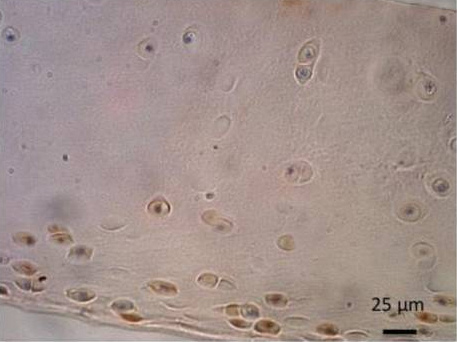
ab63084, staining TRPV6 in equine articular cartilage, by Immunohistochemistry (Formalin/PFA-fixed paraffin-embedded sections). Sections were blocked with PBS-T and 1% BSA before incubating with primary antibody overnight at 4°C. An HRP-conjugated goat anti-rabbit IgG was used as the secondary antibody and was detected using DAB. Nuclei were counterstained with hematoxylin.
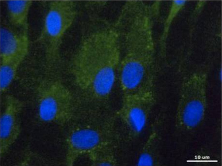
ab63084, staining TRPV6 (green) in equine articular chondrocytes, by Immunocytochemistry/ Immunofluorescence. Cells were fixed in ice-cold methanol for 10 min, washed and permeabilized with PBS-T and blocked with 10% BSA in PBS. The cells were then incubated with primary antibody overnight at 4°C. A DyLight®488-conjugated goat anti-rabbit polyclonal IgG (ab98462) was used as the secondary antibody. Nuclei were stained with DAPI (blue).