![All lanes : Anti-Transglutaminase 2 antibody [EP2957] (ab109200) at 1/10000 dilutionLane 1 : U87-MG cell lysateLane 2 : A549 cell lysateLane 3 : HUVEC cell lysate Lysates/proteins at 10 µg per lane.](http://www.bioprodhub.com/system/product_images/ab_products/2/sub_5/14014_Transglutaminase-2-Primary-antibodies-ab109200-3.jpg)
All lanes : Anti-Transglutaminase 2 antibody [EP2957] (ab109200) at 1/10000 dilutionLane 1 : U87-MG cell lysateLane 2 : A549 cell lysateLane 3 : HUVEC cell lysate Lysates/proteins at 10 µg per lane.
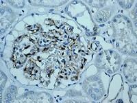
ab109200, at 1/100 dilution, staining Transglutaminase 2 in paraffin-embedded Human kidney tissue by Immunohistochemistry.
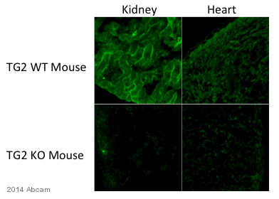
ab109200 staining Transglutaminase 2 in mouse kidney and heart tissue sections by Immunohistochemistry (IHC-Fr - frozen sections). Tissue was fixed with paraformaldehyde, permeabilized with 0.1% Tween-20 and blocked with 5% BSA for 12 hours at 4°C. Samples were incubated with primary antibody (1/500 in PBS-T + 5% BSA) for 12 hours at 4°C. An undiluted Alexa Fluor® 488-conjugated goat anti-rabbit IgG (H+L) polyclonal was used as the secondary antibody.See Abreview