![Anti-TLS/FUS antibody [EPR5813] (ab133571) at 1/1000 dilution (unpurified) + HL-60 cell lysate at 10 µgSecondaryPeroxidase-conjugated goat anti-rabbit IgG (H+L) at 1/1000 dilution](http://www.bioprodhub.com/system/product_images/ab_products/2/sub_5/11841_ab133571-241161-ab133571upwb.jpg)
Anti-TLS/FUS antibody [EPR5813] (ab133571) at 1/1000 dilution (unpurified) + HL-60 cell lysate at 10 µgSecondaryPeroxidase-conjugated goat anti-rabbit IgG (H+L) at 1/1000 dilution
![Anti-TLS/FUS antibody [EPR5813] (ab133571) at 1/2000 dilution (purified) + HL-60 cell lysate at 10 µgSecondaryPeroxidase-conjugated goat anti-rabbit IgG (H+L) at 1/1000 dilution](http://www.bioprodhub.com/system/product_images/ab_products/2/sub_5/11842_ab133571-241162-ab133571pwb.jpg)
Anti-TLS/FUS antibody [EPR5813] (ab133571) at 1/2000 dilution (purified) + HL-60 cell lysate at 10 µgSecondaryPeroxidase-conjugated goat anti-rabbit IgG (H+L) at 1/1000 dilution
![Anti-TLS/FUS antibody [EPR5813] (ab133571) at 1/1000 dilution (unpurified) + Raw264.7 cell lysate at 10 µgSecondaryPeroxidase-conjugated goat anti-rabbit IgG (H+L) at 1/1000 dilution](http://www.bioprodhub.com/system/product_images/ab_products/2/sub_5/11843_ab133571-241163-ab133571upwb2.jpg)
Anti-TLS/FUS antibody [EPR5813] (ab133571) at 1/1000 dilution (unpurified) + Raw264.7 cell lysate at 10 µgSecondaryPeroxidase-conjugated goat anti-rabbit IgG (H+L) at 1/1000 dilution
![Anti-TLS/FUS antibody [EPR5813] (ab133571) at 1/2000 dilution (purified) + Raw264.7 cell lysate at 10 µgSecondaryPeroxidase-conjugated goat anti-rabbit IgG (H+L) at 1/1000 dilution](http://www.bioprodhub.com/system/product_images/ab_products/2/sub_5/11844_ab133571-241164-ab133571pwb2.jpg)
Anti-TLS/FUS antibody [EPR5813] (ab133571) at 1/2000 dilution (purified) + Raw264.7 cell lysate at 10 µgSecondaryPeroxidase-conjugated goat anti-rabbit IgG (H+L) at 1/1000 dilution
![All lanes : Anti-TLS/FUS antibody [EPR5813] (ab133571) at 1/1000 dilution (unpurified)Lane 1 : HepG2 cell lysateLane 2 : K-562 cell lysateLane 3 : PC12 cell lysateLane 4 : NIH/3T3 cell lysateLane 5 : RAW 264.7 cell lysateLysates/proteins at 10 µg per lane.SecondaryHRP-conjugated goat anti-rabbit IgG at 1/2000 dilutiondeveloped using the ECL technique](http://www.bioprodhub.com/system/product_images/ab_products/2/sub_5/11845_TLSFUS-Primary-antibodies-ab133571-2.jpg)
All lanes : Anti-TLS/FUS antibody [EPR5813] (ab133571) at 1/1000 dilution (unpurified)Lane 1 : HepG2 cell lysateLane 2 : K-562 cell lysateLane 3 : PC12 cell lysateLane 4 : NIH/3T3 cell lysateLane 5 : RAW 264.7 cell lysateLysates/proteins at 10 µg per lane.SecondaryHRP-conjugated goat anti-rabbit IgG at 1/2000 dilutiondeveloped using the ECL technique
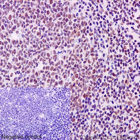
Immunohistochemistry (Formalin/PFA-fixed paraffin-embedded sections) analysis of human tonsil tissue labelling TLS/FUS with unpurified ab133571 at 1/50. Heat mediated antigen retrieval was performed using Tris/EDTA buffer pH 9. A prediluted HRP-polymer conjugated anti-rabbit IgG was used as the secondary antibody. Negative control using PBS instead of primary antibody. Counterstained with Hematoxylin.
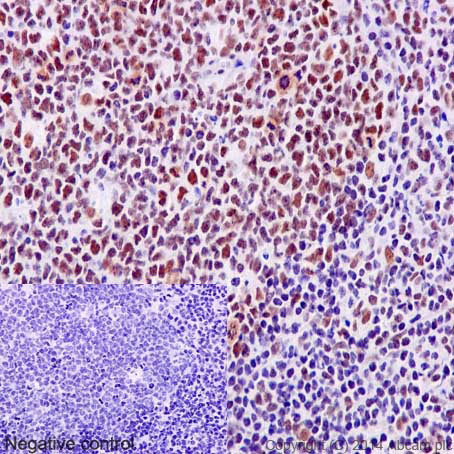
Immunohistochemistry (Formalin/PFA-fixed paraffin-embedded sections) analysis of human tonsil tissue labelling TLS/FUS with purified ab133571 at 1/100. Heat mediated antigen retrieval was performed using Tris/EDTA buffer pH 9. A prediluted HRP-polymer conjugated anti-rabbit IgG was used as the secondary antibody. Negative control using PBS instead of primary antibody. Counterstained with Hematoxylin.
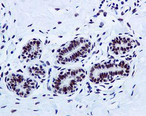
Immunohistochemistry (Formalin/PFA-fixed paraffin-embedded sections) analysis of human breast tissue labelling TLS/FUS with unpurified ab133571 at 1/100 dilution.
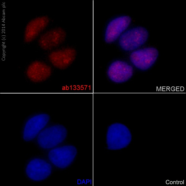
Immunocytochemistry/Immunofluorescence analysis of HeLa cells labelling TLS/FUS with purified ab133571 at 1/100. Cells were fixed with 4% paraformaldehyde. An Alexa Fluor® 555-conjugated goat anti-rabbit IgG (1/500) was used as the secondary antibody. DAPI (blue) was used as the nuclear counterstain.Control: primary antibody (1/100) and secondary antibody, ab150113, an Alexa Fluor® 488-conjugated goat anti-mouse IgG (1/500).
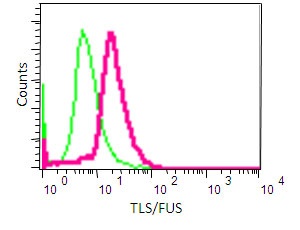
Flow cytometry analysis of NIH/3T3 cells labelling TLS/FUS with unpurified ab133571 (red) at 1/20. Cells were fixed with 2% paraformaldehyde. A FITC-conjugated goat anti-rabbit IgG (1/150) was used as the secondary antibody. Green - Isotype control, rabbit monoclonal IgG.
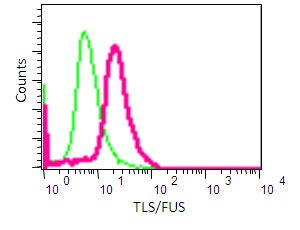
Flow cytometry analysis of NIH/3T3 cells labelling TLS/FUS with purified ab133571 (red) at 1/50. Cells were fixed with 2% paraformaldehyde. A FITC-conjugated goat anti-rabbit IgG (1/150) was used as the secondary antibody. Green - Isotype control, rabbit monoclonal IgG.
Equilibrium disassociation constant (KD)Learn more about KD Click here to learn more about KD