![All lanes : Anti-TLE 1 antibody [EPR9386(2)] (ab183742) at 1/1000 dilutionLane 1 : SH-SY5Y cell lysateLane 2 : HepG2 cell lysateLane 3 : Jurkat cell lysateLane 4 : HeLa cell lysateLysates/proteins at 20 µg per lane.SecondaryGoat Anti-Rabbit IgG, (H+L), Peroxidase conjugated at 1/1000 dilution](http://www.bioprodhub.com/system/product_images/ab_products/2/sub_5/11522_ab183742-215725-ab183742WB.jpg)
All lanes : Anti-TLE 1 antibody [EPR9386(2)] (ab183742) at 1/1000 dilutionLane 1 : SH-SY5Y cell lysateLane 2 : HepG2 cell lysateLane 3 : Jurkat cell lysateLane 4 : HeLa cell lysateLysates/proteins at 20 µg per lane.SecondaryGoat Anti-Rabbit IgG, (H+L), Peroxidase conjugated at 1/1000 dilution
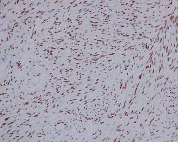
Immunohistochemical analysis of Human schwannoma, staining TLE 1 with ab183742 at 1/250 dilution. Detected using HRP Polymer for Rabbit IgG and counter-stained using hematoxylin.
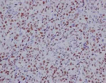
Immunohistochemical analysis of Human synovial sarcoma, staining TLE 1 with ab183742 at 1/250 dilution. Detected using HRP Polymer for Rabbit IgG and counter-stained using hematoxylin.
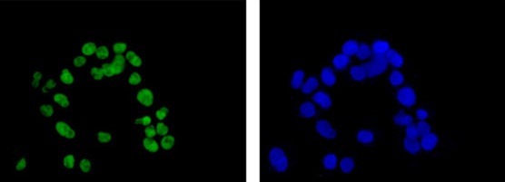
Immunofluorescence analysis of paraformaldehyde-fixed HepG2 cells, staining TLE 1 (green) with ab183742 at 1/100 dilution. Alexa Fluor®488-conjugated goat anti rabbit IgG was used as a secondary antibody at 1/200 dilution. Nuclei were counterstained with DAPI (blue).