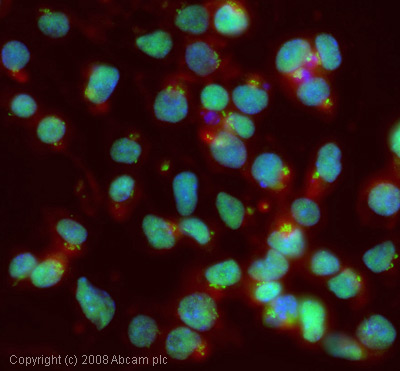
ICC/IF image of ab47062 stained mouse embryonic stem cells. The cells were 4% PFA fixed (10 min) and then incubated in 1%BSA / 10% normal goat serum / 0.3M glycine in 0.1% PBS-Tween for 1h to permeabilise the cells and block non-specific protein-protein interactions. The cells were then incubated with the antibody (ab47062, 1µg/ml) overnight at +4°C. The secondary antibody (green) was Alexa Fluor® 488 goat anti-rabbit IgG (H+L) used at a 1/1000 dilution for 1h. Alexa Fluor® 594 WGA was used to label plasma membranes (red) at a 1/200 dilution for 1h. DAPI was used to stain the cell nuclei (blue).

All lanes : Anti-TIF1 gamma antibody (ab47062) at 1 µg/mlLane 1 : F9 (Mouse embryonic carcinoma cell line) Whole Cell Lysate (ab27193)Lane 2 : E14tG2a (Mouse embryonic stem cell line) Whole Cell LysateLysates/proteins at 10 µg per lane.SecondaryIRDye 680 Conjugated Goat Anti-Rabbit IgG (H+L) at 1/10000 dilutionPerformed under reducing conditions.
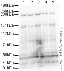
All lanes : Anti-TIF1 gamma antibody (ab47062) at 1 µg/mlLane 1 : SW480 (Human colon adenocarcinoma cell line) Whole Cell Lysate Lane 2 : HeLa (Human epithelial carcinoma cell line) Whole Cell LysateLane 3 : HEK293 (Human embryonic kidney cell line) Whole Cell LysateLane 4 : SHSY-5Y (Human neuroblastoma cell line) Whole Cell LysateLane 5 : K562 (Human erythromyeloblastoid leukemia cell line) Whole Cell LysateLysates/proteins at 10 µg per lane.SecondaryGoat Anti-Rabbit IgG H&L (HRP) preadsorbed (ab97080) at 1/5000 dilutiondeveloped using the ECL techniquePerformed under reducing conditions.