Anti-TIE2 antibody [Cl. 16]
| Name | Anti-TIE2 antibody [Cl. 16] |
|---|---|
| Supplier | Abcam |
| Catalog | ab24859 |
| Prices | $400.00 |
| Sizes | 100 µg |
| Host | Mouse |
| Clonality | Monoclonal |
| Isotype | IgG1 |
| Clone | Cl. 16 |
| Applications | IHC-F IHC-F WB ELISA FC IHC-P |
| Species Reactivities | Mouse, Human |
| Antigen | Recombinant fragment corresponding to Human TIE2 |
| Description | Mouse Monoclonal |
| Gene | TEK |
| Conjugate | Unconjugated |
| Supplier Page | Shop |
Product images
![Overlay histogram showing JEG-3 cells stained with ab24859 (red line). The cells were fixed with methanol (5 min) and incubated in 1x PBS / 10% normal goat serum / 0.3M glycine to block non-specific protein-protein interactions. The cells were then incubated with the antibody (ab24859, 2µg/1x106 cells) for 30 min at 22°C. The secondary antibody used was DyLight® 488 goat anti-mouse IgG (H+L) (ab96879) at 1/500 dilution for 30 min at 22°C. Isotype control antibody (black line) was mouse IgG1 [ICIGG1] (ab91353, 2µg/1x106 cells) used under the same conditions. Acquisition of >5,000 events was performed. This antibody gave a decreased signal in JEG-3 cells fixed with 4% paraformaldehyde (10 min) used under the same conditions.Please note that Abcam does not have data for use of this antibody on non-fixed cells. We welcome any customer feedback.](http://www.bioprodhub.com/system/product_images/ab_products/2/sub_5/11075_TIE2-Primary-antibodies-ab24859-2.jpg) Overlay histogram showing JEG-3 cells stained with ab24859 (red line). The cells were fixed with methanol (5 min) and incubated in 1x PBS / 10% normal goat serum / 0.3M glycine to block non-specific protein-protein interactions. The cells were then incubated with the antibody (ab24859, 2µg/1x106 cells) for 30 min at 22°C. The secondary antibody used was DyLight® 488 goat anti-mouse IgG (H+L) (ab96879) at 1/500 dilution for 30 min at 22°C. Isotype control antibody (black line) was mouse IgG1 [ICIGG1] (ab91353, 2µg/1x106 cells) used under the same conditions. Acquisition of >5,000 events was performed. This antibody gave a decreased signal in JEG-3 cells fixed with 4% paraformaldehyde (10 min) used under the same conditions.Please note that Abcam does not have data for use of this antibody on non-fixed cells. We welcome any customer feedback.
Overlay histogram showing JEG-3 cells stained with ab24859 (red line). The cells were fixed with methanol (5 min) and incubated in 1x PBS / 10% normal goat serum / 0.3M glycine to block non-specific protein-protein interactions. The cells were then incubated with the antibody (ab24859, 2µg/1x106 cells) for 30 min at 22°C. The secondary antibody used was DyLight® 488 goat anti-mouse IgG (H+L) (ab96879) at 1/500 dilution for 30 min at 22°C. Isotype control antibody (black line) was mouse IgG1 [ICIGG1] (ab91353, 2µg/1x106 cells) used under the same conditions. Acquisition of >5,000 events was performed. This antibody gave a decreased signal in JEG-3 cells fixed with 4% paraformaldehyde (10 min) used under the same conditions.Please note that Abcam does not have data for use of this antibody on non-fixed cells. We welcome any customer feedback.
 Immunohistochemical analysis of Human brain tissue, staining TIE2 with ab24859.Tissue was fixed with paraformaldehyde, permeabilized with 0.25 Triton X-100 and blocked with 2.5% BSA for 30 minutes at 25°C. Samples were incubated with primary antibody (1/200 in 2.5% horse serum) for 18 hours at 4°C. An HRP-conjugated horse anti-rabbit polyclonal IgG was used as the secondary antibody. See Abreview
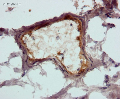
 ab24859 staining TIE2 in Human spleen by Immunohistochemistry (Frozen sections).
![All lanes : Anti-TIE2 antibody [Cl. 16] (ab24859)Lane 1 : HUVECs left untreatedLane 2 : HUVECs stimulated for 3 hours with PMA at 25 ng/mlLane 3 : HUVECs stimulated for 6 hours with PMA at 25 ng/mlLane 4 : HUVECs stimulated for 9 hours with PMA at 25 ng/mlLane 5 : HUVECs stimulated for 24 hours with PMA at 25 ng/ml](http://www.bioprodhub.com/system/product_images/ab_products/2/sub_5/11078_TIE2-Primary-antibodies-ab24859-4.jpg) All lanes : Anti-TIE2 antibody [Cl. 16] (ab24859)Lane 1 : HUVECs left untreatedLane 2 : HUVECs stimulated for 3 hours with PMA at 25 ng/mlLane 3 : HUVECs stimulated for 6 hours with PMA at 25 ng/mlLane 4 : HUVECs stimulated for 9 hours with PMA at 25 ng/mlLane 5 : HUVECs stimulated for 24 hours with PMA at 25 ng/ml
All lanes : Anti-TIE2 antibody [Cl. 16] (ab24859)Lane 1 : HUVECs left untreatedLane 2 : HUVECs stimulated for 3 hours with PMA at 25 ng/mlLane 3 : HUVECs stimulated for 6 hours with PMA at 25 ng/mlLane 4 : HUVECs stimulated for 9 hours with PMA at 25 ng/mlLane 5 : HUVECs stimulated for 24 hours with PMA at 25 ng/ml
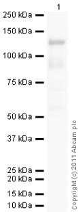 developed using the ECL techniquePerformed under reducing conditions.
Product References
Targeting of beta adrenergic receptors results in therapeutic efficacy against - Targeting of beta adrenergic receptors results in therapeutic efficacy against
Stiles JM, Amaya C, Rains S, Diaz D, Pham R, Battiste J, Modiano JF, Kokta V, Boucheron LE, Mitchell DC, Bryan BA. PLoS One. 2013;8(3):e60021.
Telomerase-based immortalization modifies the angiogenic/inflammatory responses - Telomerase-based immortalization modifies the angiogenic/inflammatory responses
Baumer Y, Scholz B, Ivanov S, Schlosshauer B. Exp Biol Med (Maywood). 2011 Jun 1;236(6):692-700.
Control of HIF-1{alpha} and vascular signaling in fetal lung involves cross talk - Control of HIF-1{alpha} and vascular signaling in fetal lung involves cross talk
Scott CL, Walker DJ, Cwiklinski E, Tait C, Tee AR, Land SC. Am J Physiol Lung Cell Mol Physiol. 2010 Oct;299(4):L455-71. doi:
CD133-expressing stem cells associated with ovarian metastases establish an - CD133-expressing stem cells associated with ovarian metastases establish an
Kusumbe AP, Mali AM, Bapat SA. Stem Cells. 2009 Mar;27(3):498-508.