Anti-TGF beta induced factor 2 antibody - C-terminal
| Name | Anti-TGF beta induced factor 2 antibody - C-terminal |
|---|---|
| Supplier | Abcam |
| Catalog | ab190152 |
| Prices | $407.00 |
| Sizes | 200 µl |
| Host | Rabbit |
| Clonality | Polyclonal |
| Isotype | IgG |
| Applications | FC WB IHC-P |
| Species Reactivities | Mouse, Human |
| Antigen | Synthetic peptide corresponding to Human TGF beta induced factor 2 aa 170-199 (C terminal) conjugated to Keyhole Limpet Haemocyanin (KLH) |
| Description | Rabbit Polyclonal |
| Gene | TGIF2 |
| Conjugate | Unconjugated |
| Supplier Page | Shop |
Product images
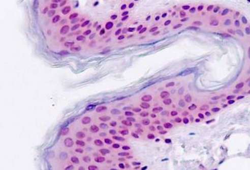 Immunohistochemical analysis of formalin-fixed, paraffin-embedded Human skin tissue labeling TGF beta induced factor 2 with ab190152 at 5 µg/ml.
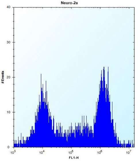 Flow cytometric analysis of Neuro-2a cells (right histogram) compared to a negative control cell (left histogram). TGF beta induced factor 2 was labeled using ab190152 at 1/50 dilution, FITC-conjugated donkey-anti-rabbit secondary antibodies were used for the analysis.
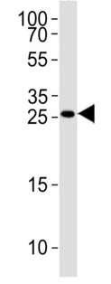 Anti-TGF beta induced factor 2 antibody - C-terminal (ab190152) at 1/1000 dilution + 293 cell line lysates at 35 µg