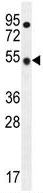
Anti-TBC1D13 antibody (ab170529) at 1/100 dilution + CEM cell line lysates at 35 µgdeveloped using the ECL technique
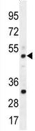
Anti-TBC1D13 antibody (ab170529) at 1/100 dilution + Mouse spleen tissue lysate at 35 µgdeveloped using the ECL technique
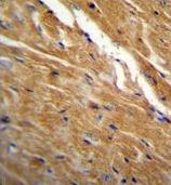
Immunohistochemical analysis of formalin-fixed, paraffin-embedded Mouse heart tissue labeling TBC1D13 with ab170529 at 1/50 dilution followed by peroxidase-conjugated secondary antibody and DAB staining.
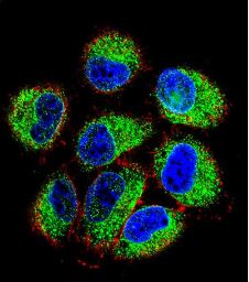
Immunofluorescence analysis of NCI-H460 cells labeling TBC1D13 with ab170529 at 1/10 dilution, followed by Alexa Fluor 488-conjugated Goat anti-Rabbit lgG (green). Actin filaments have been labeled with Alexa Fluor 555 phalloidin (red). DAPI was used to stain the cell nuclear (blue).