Anti-TAF11 antibody
| Name | Anti-TAF11 antibody |
|---|---|
| Supplier | Abcam |
| Catalog | ab57501 |
| Prices | $370.00 |
| Sizes | 100 µg |
| Host | Mouse |
| Clonality | Monoclonal |
| Isotype | IgG1 |
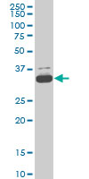
| Applications | WB IHC-P |
| Species Reactivities | Mouse, Human |
| Antigen | Recombinant fragment: SKVFVGEVVE EALDVCEKWG EMPPLQPKHM REAVRRLKSK GQIPNSKHKK IIF, corresponding to amino acids 158-211 of Human TAF11 Run BLAST with Run BLAST with |
| Description | Mouse Monoclonal |
| Gene | TAF11 |
| Conjugate | Unconjugated |
| Supplier Page | Shop |
Product images
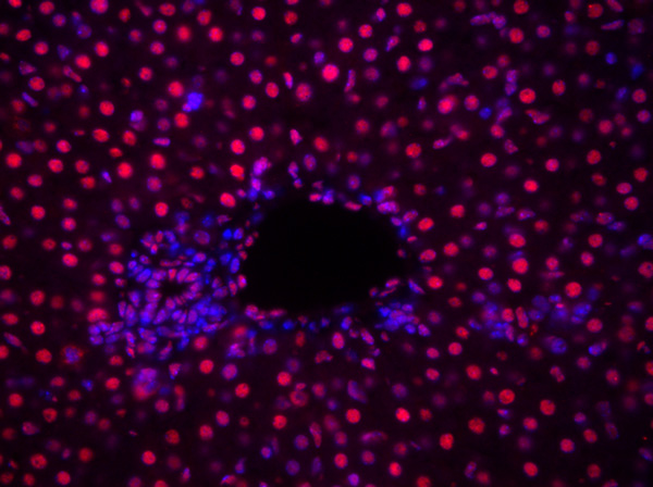 ab57501 staining TAF11 in mouse liver tissue sections by Immunohistochemistry (IHC-P - paraformaldehyde-fixed, paraffin-embedded sections). Tissue was fixed with paraformaldehyde, permeabilized with 0.1% Triton in PBS for 10 minutes and blocked with 5% BSA for 1 hour at 25°C; antigen retrieval was by heat mediation in sodium citrate buffer, pH 6. Samples were incubated with primary antibody (1/500 in 0.5% Triton + PBS) for 16 hours at 4°C. A Cy3®-conjugated goat anti-mouse IgG polyclonal (1/400) was used as the secondary antibody.See Abreview
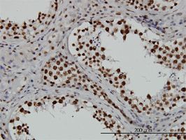 TAF11 antibody (ab57501) used in immunohistochemistry at 3ug/ml on formalin fixed and paraffin embedded human testis.