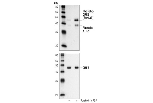
Western blot analysis of extracts from SK-N-MC cells, untreated or forskolin- and FGF-treated, using Phospho-CREB (Ser133) (87G3) Rabbit mAb (upper) or CREB (48H2) Rabbit mAb #9197 (lower).
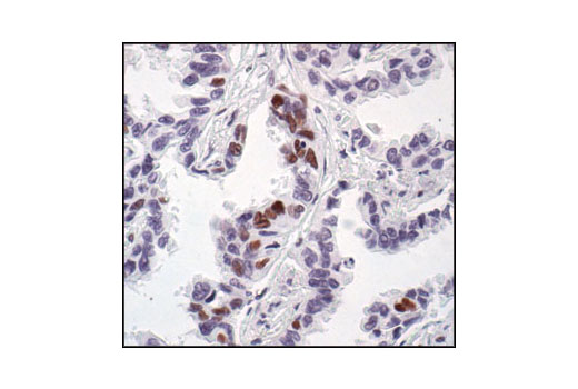
Immunohistochemical analysis of paraffin-embedded human lung carcinoma, showing nuclear staining, using Phospho-CREB (Ser133) (87G3) Rabbit mAb.
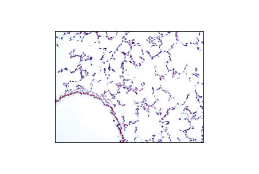
Immunohistochemical analysis of paraffin-embedded mouse lung using Phospho-CREB (Ser133) (87G3) Rabbit mAb.
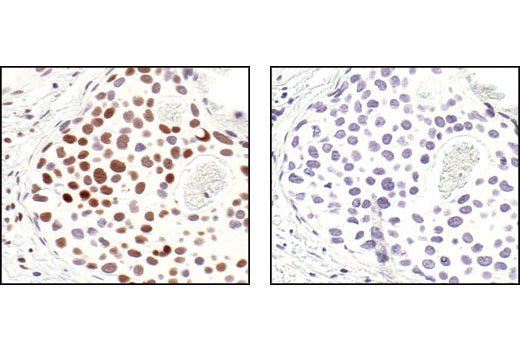
Immunohistochemical analysis of paraffin-embedded human breast carcinoma, using Phospho-CREB (Ser133) (87G3) Rabbit mAb in the presence of control peptide (left) or Phospho-CREB (Ser133) Blocking Peptide #1090 (right).
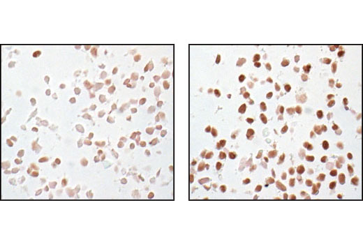
Immunohistochemical analysis of paraffin-embedded SK-N-MC cells, untreated (left) or IBMX- and forskolin-treated (right), showing induced nuclear staining, using Phospho-CREB (Ser133) (87G3) Rabbit mAb.
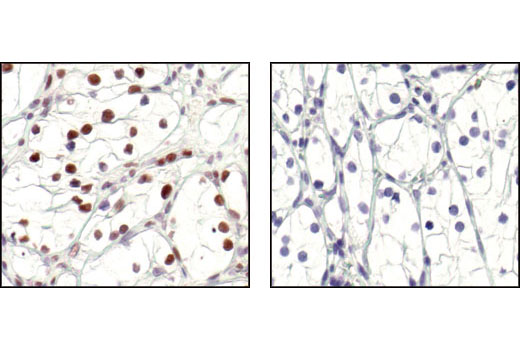
Immunohistochemical analysis of paraffin-embedded human renal cell carcinoma, untreated (left) or lambda phosphatase-treated (right), using Phospho-CREB (Ser133) (87G3) Rabbit mAb.
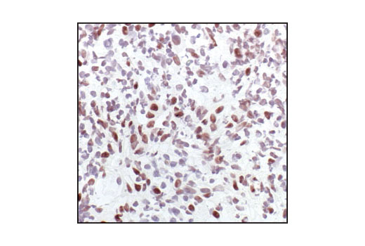
Immunohistochemical analysis of frozen H1650 xenograft, showing nuclear localization using Phospho-CREB (Ser133)(87G3) Rabbit mAb.
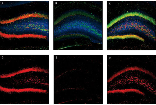
Conofocal immunofluorescent images of rat dentate gyrus, either sham-operated (left) or 15 min ischemia followed by 30 min (center) and 4 h (right) reperfusion, labeled with Phospho-CREB (Ser133) (87G3) Rabbit mAb (red), Neurofilament-L (DA2) Mouse mAb #2835 (blue) and Phospho-S6 Ribosomal Protein (Ser235/236) (2F9) Rabbit mAb (Alexa Fluor® 488 Conjugate) #4854.
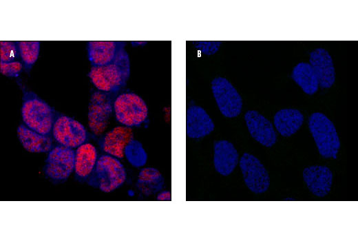
Confocal microscopic images of SK-N-MC cells showing nuclear stain after 25 minute treatment with Forskolin and IBMX using Phospho-CREB (Ser133) (87G3) Rabbit mAb (left, red) compared to untreated cells (right). Blue pseudocolor = DRAQ5 ® #4084 (fluorescent DNA dye).
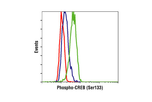
Flow cytometric analysis of SK-N-MC cells, untreated (blue) or IBMX- and forskolin-treated (green), using Phospho-CREB (Ser133) (87G3) Rabbit mAb compared to a nonspecific negative control antibody (red).
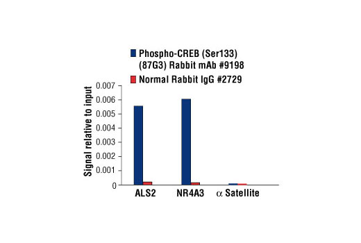
Chromatin immunoprecipitations were performed with cross-linked chromatin from 4 x 10 6 293 cells treated with Forskolin #3828 (30 μM) for 1h and either 10 μl of Phospho-CREB (Ser133) (87G3) Rabbit mAb or 2 μl of Normal Rabbit IgG #2729 using SimpleChIP ® Enzymatic Chromatin IP Kit (Magnetic Beads) #9003. The enriched DNA was quantified by real-time PCR using human ALS2 exon 1 primers, SimpleChIP ® Human NR4A3 Promoter Primers #4829, and SimpleChIP ® Human α Satellite Repeat Primers #4486. The amount of immunoprecipitated DNA in each sample is represented as signal relative to the total amount of input chromatin, which is equivalent to one.


