![All lanes : Anti-SP1 antibody [EPR6661] (ab133596) at 1/10000 dilution (purified)Lane 1 : A431 cell lysateLane 2 : HeLa cell lysateLane 3 : A549 cell lysateLysates/proteins at 20 µg per lane.SecondaryHRP goat anti-rabbit (H+L) at 1/1000 dilution](http://www.bioprodhub.com/system/product_images/ab_products/2/sub_5/3933_ab133596-239899-133596-WB-2.jpg)
All lanes : Anti-SP1 antibody [EPR6661] (ab133596) at 1/10000 dilution (purified)Lane 1 : A431 cell lysateLane 2 : HeLa cell lysateLane 3 : A549 cell lysateLysates/proteins at 20 µg per lane.SecondaryHRP goat anti-rabbit (H+L) at 1/1000 dilution
![All lanes : Anti-SP1 antibody [EPR6661] (ab133596) at 1/2000 dilution (purified)Lane 1 : Jurkat cell lysateLane 2 : K562 cell lysateSecondaryHRP goat anti-rabbit (H+L) at 1/1000 dilution](http://www.bioprodhub.com/system/product_images/ab_products/2/sub_5/3934_ab133596-239896-133596-WB-1.jpg)
All lanes : Anti-SP1 antibody [EPR6661] (ab133596) at 1/2000 dilution (purified)Lane 1 : Jurkat cell lysateLane 2 : K562 cell lysateSecondaryHRP goat anti-rabbit (H+L) at 1/1000 dilution
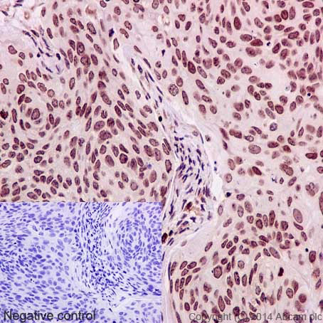
Immunohistochemical staining of paraffin embedded human cervical carcinoma with purified ab133596 at a working dilution of 1 in 250. The secondary antibody used is a HRP polymer for rabbit IgG. The sample is counter-stained with hematoxylin. Antigen retrieval was perfomed using Tris-EDTA buffer, pH 9.0. PBS was used instead of the primary antibody as the negative control, and is shown in the inset.
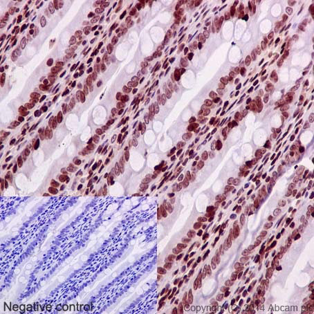
Immunohistochemical staining of paraffin embedded rat colon with purified ab133596 at a working dilution of 1 in 250. The secondary antibody used is a HRP polymer for rabbit IgG. The sample is counter-stained with hematoxylin. Antigen retrieval was perfomed using Tris-EDTA buffer, pH 9.0. PBS was used instead of the primary antibody as the negative control, and is shown in the inset.
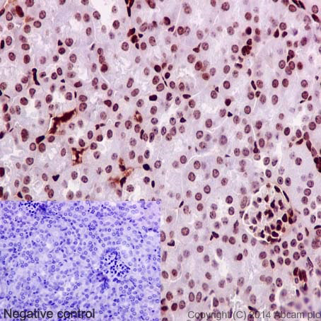
Immunohistochemical staining of paraffin embedded mouse kidney with purified ab133596 at a working dilution of 1 in 250. The secondary antibody used is a HRP polymer for rabbit IgG. The sample is counter-stained with hematoxylin. Antigen retrieval was perfomed using Tris-EDTA buffer, pH 9.0. PBS was used instead of the primary antibody as the negative control, and is shown in the inset.
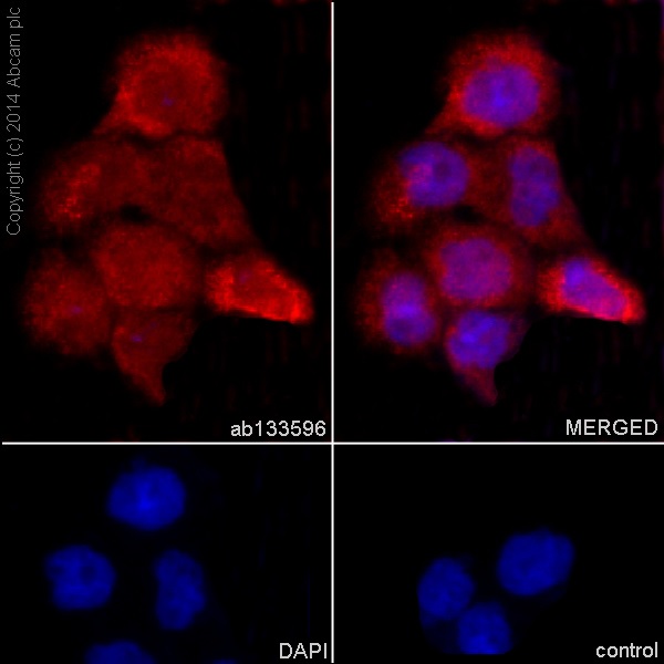
Immunofluorescence staining of A431 cells with purified ab133596 at a working dilution of 1 in 250, counter-stained with DAPI. The secondary antibody was Alexa Fluor® 555 goat anti rabbit, used at a dilution of 1 in 500. The cells were fixed in 4% PFA and permeabilized using 0.1% Triton X 100. The negative control is shown in bottom right hand panel - for the negative control, purified ab133596 was used at a dilution of 1/200 followed by an Alexa Fluor® 488 goat anti-mouse antibody (ab150113) at a dilution of 1/500.
![All lanes : Anti-SP1 antibody [EPR6661] (ab133596) at 1/1000 dilution (unpurified)Lane 1 : A431 cell lysateLane 2 : HeLa cell lysateLane 3 : A549 cell lysateLane 4 : K562 cell lysateLane 5 : Jurkat cell lysateLysates/proteins at 10 µg per lane.SecondaryHRP labelled goat anti-rabbit at 1/2000 dilution](http://www.bioprodhub.com/system/product_images/ab_products/2/sub_5/3939_SP1-Primary-antibodies-ab133596-1.jpg)
All lanes : Anti-SP1 antibody [EPR6661] (ab133596) at 1/1000 dilution (unpurified)Lane 1 : A431 cell lysateLane 2 : HeLa cell lysateLane 3 : A549 cell lysateLane 4 : K562 cell lysateLane 5 : Jurkat cell lysateLysates/proteins at 10 µg per lane.SecondaryHRP labelled goat anti-rabbit at 1/2000 dilution
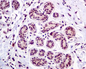
Immunohistochemical analysis of paraffin-embedded Human breast tissue labelling SP1 with unpurified ab133596 at 1/100 dilution.
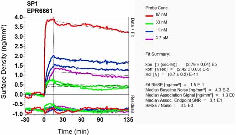
Equilibrium disassociation constant (KD)Learn more about KD Click here to learn more about KD