Anti-SP1 antibody
| Name | Anti-SP1 antibody |
|---|---|
| Supplier | Abcam |
| Catalog | ab77441 |
| Prices | $398.00 |
| Sizes | 100 µg |
| Host | Mouse |
| Clonality | Monoclonal |
| Isotype | IgG1 |
| Applications | WB ELISA IHC-P ICC/IF ICC/IF FC |
| Species Reactivities | Mouse, Human |
| Antigen | Recombinant fragment, corresponding to amino acids 89-199 of Human SP1 with tag (NP_612482) |
| Description | Mouse Monoclonal |
| Gene | SP1 |
| Conjugate | Unconjugated |
| Supplier Page | Shop |
Product images
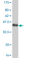 Anti-SP1 antibody (ab77441) at 5 µg/ml + Recombinant fragment of human SP1 at 0.2 µgSecondaryGoat anti-Mouse IgG (H&L)-HRP at 1/5000 dilution
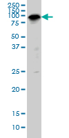 Anti-SP1 antibody (ab77441) at 5 µg/ml + IMR-32 cell lysate at 25 µgSecondaryGoat anti-Mouse IgG (H&L)-HRP at 1/2500 dilution
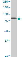 Anti-SP1 antibody (ab77441) at 5 µg/ml + HeLa nuclear cell lysate at 25 µgSecondaryGoat anti-Mouse IgG (H&L)-HRP at 1/2500 dilution
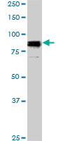 Anti-SP1 antibody (ab77441) at 5 µg/ml + NIH3T3 cell lysate at 25 µgSecondaryGoat anti-Mouse IgG (H&L)-HRP at 1/2500 dilution
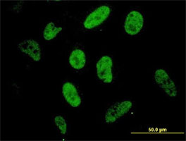 ab77441 at 10µg/ml staining SP1 in HeLa cells by Immunofluorescence.
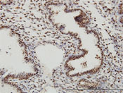 ab77441 staining SP1 in human endometrium by Immunohistochemistry, Formalin-fixed, Paraffin-embedded tissue.
![Overlay histogram showing HeLa cells stained with ab77441 (red line). The cells were fixed with methanol (5 min) and then permeabilized with 0.1% PBS-Tween for 20 min. The cells were then incubated in 1x PBS / 10% normal goat serum / 0.3M glycine to block non-specific protein-protein interactions followed by the antibody (ab77441, 2µg/1x106 cells) for 30 min at 22°C. The secondary antibody used was DyLight® 488 goat anti-mouse IgG (H+L) (ab96879) at 1/500 dilution for 30 min at 22°C. Isotype control antibody (black line) was mouse mouse IgG1 [ICIGG1] (ab91353, 2µg/1x106 cells) used under the same conditions. Acquisition of >5,000 events was performed. This antibody gave a diminished signal in HeLa cells fixed with 4% paraformaldehyde (10 min)/permeabilized with 0.1% PBS-Tween 20 used under the same conditions.](http://www.bioprodhub.com/system/product_images/ab_products/2/sub_5/3910_SP1-Primary-antibodies-ab77441-4.jpg) Overlay histogram showing HeLa cells stained with ab77441 (red line). The cells were fixed with methanol (5 min) and then permeabilized with 0.1% PBS-Tween for 20 min. The cells were then incubated in 1x PBS / 10% normal goat serum / 0.3M glycine to block non-specific protein-protein interactions followed by the antibody (ab77441, 2µg/1x106 cells) for 30 min at 22°C. The secondary antibody used was DyLight® 488 goat anti-mouse IgG (H+L) (ab96879) at 1/500 dilution for 30 min at 22°C. Isotype control antibody (black line) was mouse mouse IgG1 [ICIGG1] (ab91353, 2µg/1x106 cells) used under the same conditions. Acquisition of >5,000 events was performed. This antibody gave a diminished signal in HeLa cells fixed with 4% paraformaldehyde (10 min)/permeabilized with 0.1% PBS-Tween 20 used under the same conditions.
Overlay histogram showing HeLa cells stained with ab77441 (red line). The cells were fixed with methanol (5 min) and then permeabilized with 0.1% PBS-Tween for 20 min. The cells were then incubated in 1x PBS / 10% normal goat serum / 0.3M glycine to block non-specific protein-protein interactions followed by the antibody (ab77441, 2µg/1x106 cells) for 30 min at 22°C. The secondary antibody used was DyLight® 488 goat anti-mouse IgG (H+L) (ab96879) at 1/500 dilution for 30 min at 22°C. Isotype control antibody (black line) was mouse mouse IgG1 [ICIGG1] (ab91353, 2µg/1x106 cells) used under the same conditions. Acquisition of >5,000 events was performed. This antibody gave a diminished signal in HeLa cells fixed with 4% paraformaldehyde (10 min)/permeabilized with 0.1% PBS-Tween 20 used under the same conditions.
Product References
Influenza polymerase encoding mRNAs utilize atypical mRNA nuclear export. - Influenza polymerase encoding mRNAs utilize atypical mRNA nuclear export.
Larsen S, Bui S, Perez V, Mohammad A, Medina-Ramirez H, Newcomb LL. Virol J. 2014 Aug 28;11:154.
Divergent Sp1 protein levels may underlie differential expression of UDP-glucose - Divergent Sp1 protein levels may underlie differential expression of UDP-glucose
Tsui S, Fernando R, Chen B, Smith TJ. J Biol Chem. 2011 Jul 8;286(27):24487-99.