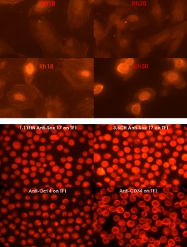
Top image: Immunofluorescence staining of Rh18 and Rh30 Cells after treatment with Aphidicolin or Nocadazole labeling SOX17 using ab192453 and PE anti-mouse Ig labeled secondary antibody.Bottom image: Immunofluorescence staining of TF1 hematopoietic stem cell line labeling SOX17 using ab192453 and TRITC labeled anti-mouse Ig secondary antibody.
![Anti-SOX17 [BC24-3.5CH] antibody (ab192453) at 2 µg/ml + MDCK cell lysate at 20 µgSecondaryPeroxidase conjugated rabbit anti-mouse Ig at 1/20000 dilution](http://www.bioprodhub.com/system/product_images/ab_products/2/sub_5/3618_ab192453-229372-ab192453-3.jpg)
Anti-SOX17 [BC24-3.5CH] antibody (ab192453) at 2 µg/ml + MDCK cell lysate at 20 µgSecondaryPeroxidase conjugated rabbit anti-mouse Ig at 1/20000 dilution
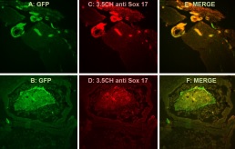
Immunofluorescence analysis of endothelial cells and gut endoderm, staining SOX17 using ab192453 and TRITC labeled anti mouse Ig secondary antibody.
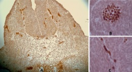
Immunohistochemical analysis of PFA-fixed mouse embryo gut endoderm and endothelial cells, labeling SOX17 using ab192453 at 5 ug/mL and peroxidase labeled anti-mouse Ig secondary antibody, with DAB staining.