Anti-SMARCA1 antibody - ChIP Grade
| Name | Anti-SMARCA1 antibody - ChIP Grade |
|---|---|
| Supplier | Abcam |
| Catalog | ab37003 |
| Prices | $370.00 |
| Sizes | 100 µg |
| Host | Rabbit |
| Clonality | Polyclonal |
| Isotype | IgG |
| Applications | IP WB ICC/IF ICC/IF ChIP |
| Species Reactivities | Human, Mouse, Drosophila, Zebrafish |
| Antigen | Synthetic peptide conjugated to KLH derived from within residues 850 - 950 of Human SMARCA1 |
| Blocking Peptide | Human SMARCA1 peptide |
| Description | Rabbit Polyclonal |
| Gene | SMARCA1 |
| Conjugate | Unconjugated |
| Supplier Page | Shop |
Product images
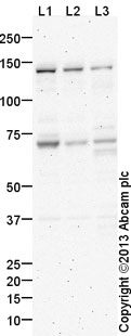 All lanes : Anti-SMARCA1 antibody - ChIP Grade (ab37003) at 1 µg/ml (blocked with 3% milk)Lane 1 : Jurkat (Human T cell lymphoblast-like cell line) Nuclear Lysate (ab14844)Lane 2 : HEK293 (Human embryonic kidney cell line) Nuclear LysateLane 3 : SHSY5Y Whole Cell LysateLysates/proteins at 20 µg per lane.SecondaryGoat Anti-Rabbit IgG H&L (HRP) (ab97051) at 1/10000 dilutiondeveloped using the ECL techniquePerformed under reducing conditions.
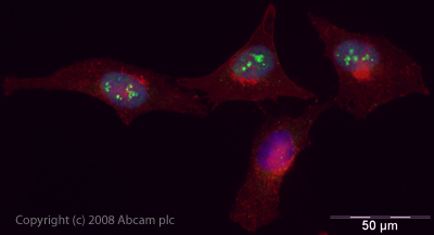 ICC/IF image of ab37003 stained human HeLa cells. The cells were PFA fixed (10 min), permabilised in PBS-T (20 min) and incubated with the antibody (ab37003, 5µg/ml) for 1h at room temperature. 1%BSA / 10% normal goat serum / 0.3M glycine was used to quench autofluorescence and block non-specific protein-protein interactions. The secondary antibody (green) was Alexa Fluor® 488 goat anti-rabbit IgG (H+L) used at a 1/1000 dilution for 1h. Alexa Fluor® 594 WGA was used to label plasma membranes (red). DAPI was used to stain the cell nuclei (blue).
![SMARCA1 was immunoprecipitated using 0.5mg Jurkat whole cell extract, 5µg of Rabbit polyclonal to SMARCA1 and 50µl of protein G magnetic beads (+). No antibody was added to the control (-). The antibody was incubated under agitation with Protein G beads for 10min, Jurkat whole cell extract lysate diluted in RIPA buffer was added to each sample and incubated for a further 10min under agitation.Proteins were eluted by addition of 40µl SDS loading buffer and incubated for 10min at 70oC; 10µl of each sample was separated on a SDS PAGE gel, transferred to a nitrocellulose membrane, blocked with 5% BSA and probed with ab37003.Secondary: Mouse monoclonal [SB62a] Secondary Antibody to Rabbit IgG light chain (HRP) (ab99697).Band: 105kDa: SMARCA1.](http://www.bioprodhub.com/system/product_images/ab_products/2/sub_5/2032_SMARCA1-Primary-antibodies-ab37003-3.jpg) SMARCA1 was immunoprecipitated using 0.5mg Jurkat whole cell extract, 5µg of Rabbit polyclonal to SMARCA1 and 50µl of protein G magnetic beads (+). No antibody was added to the control (-). The antibody was incubated under agitation with Protein G beads for 10min, Jurkat whole cell extract lysate diluted in RIPA buffer was added to each sample and incubated for a further 10min under agitation.Proteins were eluted by addition of 40µl SDS loading buffer and incubated for 10min at 70oC; 10µl of each sample was separated on a SDS PAGE gel, transferred to a nitrocellulose membrane, blocked with 5% BSA and probed with ab37003.Secondary: Mouse monoclonal [SB62a] Secondary Antibody to Rabbit IgG light chain (HRP) (ab99697).Band: 105kDa: SMARCA1.
SMARCA1 was immunoprecipitated using 0.5mg Jurkat whole cell extract, 5µg of Rabbit polyclonal to SMARCA1 and 50µl of protein G magnetic beads (+). No antibody was added to the control (-). The antibody was incubated under agitation with Protein G beads for 10min, Jurkat whole cell extract lysate diluted in RIPA buffer was added to each sample and incubated for a further 10min under agitation.Proteins were eluted by addition of 40µl SDS loading buffer and incubated for 10min at 70oC; 10µl of each sample was separated on a SDS PAGE gel, transferred to a nitrocellulose membrane, blocked with 5% BSA and probed with ab37003.Secondary: Mouse monoclonal [SB62a] Secondary Antibody to Rabbit IgG light chain (HRP) (ab99697).Band: 105kDa: SMARCA1.
Product References
Multiple epigenetic modifiers induce aggressive viral extinction in - Multiple epigenetic modifiers induce aggressive viral extinction in
Golding MC, Zhang L, Mann MR. Cell Stem Cell. 2010 May 7;6(5):457-67.
Inhibition of expression of the chromatin remodeling gene, SNF2L, selectively - Inhibition of expression of the chromatin remodeling gene, SNF2L, selectively
Ye Y, Xiao Y, Wang W, Wang Q, Yearsley K, Wani AA, Yan Q, Gao JX, Shetuni BS, Barsky SH. Mol Cancer Res. 2009 Dec;7(12):1984-99.