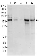
All lanes : Anti-SIK3 antibody (ab88495) at 0.4 µg/mlLane 1 : HeLa cell lysate at 50 µgLane 2 : HeLa cell lysate at 15 µgLane 3 : HeLa cell lysate at 5 µgLane 4 : 293T cell lysate at 50 µgLane 5 : NIH3T3 cell lysate at 50 µg
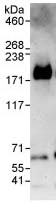
3ug ab88495 were used to immunoprecipitate 1mg HeLa whole cell lysate. 20% of immunoprecipitate was loaded on gel and probed with ab88495 at 1ug/ml. Detection: chemoluminescence with exposure time of 3 minutes.
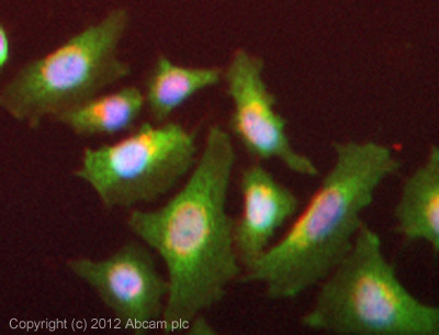
ICC/IF image of ab88495 stained HeLa cells. The cells were 4% formaldehyde (10 min) and then incubated in 1%BSA / 10% normal goat serum / 0.3M glycine in 0.1% PBS-Tween for 1h to permeabilise the cells and block non-specific protein-protein interactions. The cells were then incubated with the antibody (ab88495, 5µg/ml) overnight at +4°C. The secondary antibody (green) was ab96899 Dylight 488 goat anti-rabbit IgG (H+L) used at a 1/250 dilution for 1h. Alexa Fluor® 594 WGA was used to label plasma membranes (red) at a 1/200 dilution for 1h. DAPI was used to stain the cell nuclei (blue) at a concentration of 1.43µM.