Anti-SHMT1 antibody
| Name | Anti-SHMT1 antibody |
|---|---|
| Supplier | Abcam |
| Catalog | ab186130 |
| Prices | $370.00 |
| Sizes | 100 µg |
| Host | Rabbit |
| Clonality | Polyclonal |
| Isotype | IgG |
| Applications | IP WB |
| Species Reactivities | Mouse, Human, Dog, Chimpanzee, Monkey, Monkey, Ape, Orangutan |
| Antigen | Synthetic peptide within Human SHMT1 aa 453-483 (C terminal) |
| Description | Rabbit Polyclonal |
| Gene | SHMT1 |
| Conjugate | Unconjugated |
| Supplier Page | Shop |
Product images
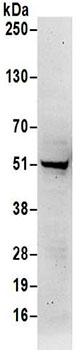 Anti-SHMT1 antibody (ab186130) at 1 µg/ml + Jurkat whole cell lysate at 50 µgdeveloped using the ECL technique
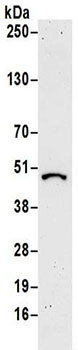 Anti-SHMT1 antibody (ab186130) at 1 µg/ml + NIH 3T3 whole cell lysate at 50 µgdeveloped using the ECL technique
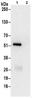 Detection of SHMT1 in immunoprecipitates of Jurkat whole cell lysate (1 mg for IP, 20% of IP loaded) using ab186130 at 6 µg per reaction for IP (Lane 1) and at 1 µg/ml for subsequent Western blot detection. Lane 2 represents control IgG IP.Detection: Chemiluminescence with an exposure time of 30 seconds.