![Anti-SGTB antibody [EPR17183] (ab202419) at 1/2000 dilution + HepG2 (Human liver hepatocellular carcinoma) cell lysate at 20 µgSecondaryGoat Anti-Rabbit IgG, (H+L), Peroxidase conjugated at 1/1000 dilution](http://www.bioprodhub.com/system/product_images/ab_products/2/sub_4/29390_ab202419-245008-ab202419-wb-1.jpg)
Anti-SGTB antibody [EPR17183] (ab202419) at 1/2000 dilution + HepG2 (Human liver hepatocellular carcinoma) cell lysate at 20 µgSecondaryGoat Anti-Rabbit IgG, (H+L), Peroxidase conjugated at 1/1000 dilution
![Anti-SGTB antibody [EPR17183] (ab202419) at 1/2000 dilution + Human fetal brain lysate at 10 µgSecondaryGoat Anti-Rabbit IgG, (H+L), Peroxidase conjugated at 1/1000 dilution](http://www.bioprodhub.com/system/product_images/ab_products/2/sub_4/29391_ab202419-245007-ab202419-wb-2.jpg)
Anti-SGTB antibody [EPR17183] (ab202419) at 1/2000 dilution + Human fetal brain lysate at 10 µgSecondaryGoat Anti-Rabbit IgG, (H+L), Peroxidase conjugated at 1/1000 dilution
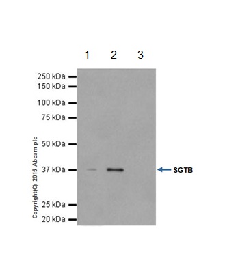
![All lanes : Anti-SGTB antibody [EPR17183] (ab202419) at 1/2000 dilutionLane 1 : Mouse brain lysateLane 2 : Rat brain lysateLane 3 : PC-12 (Rat adrenal gland pheochromocytoma) cell lysateLane 4 : NIH/3T3 (Mouse embyro fibroblast cells) cell lysateLysates/proteins at 10 µg per lane.SecondaryGoat Anti-Rabbit IgG, (H+L), Peroxidase conjugated at 1/1000 dilution](http://www.bioprodhub.com/system/product_images/ab_products/2/sub_4/29392_ab202419-245006-ab202419-wb-3.jpg)
All lanes : Anti-SGTB antibody [EPR17183] (ab202419) at 1/2000 dilutionLane 1 : Mouse brain lysateLane 2 : Rat brain lysateLane 3 : PC-12 (Rat adrenal gland pheochromocytoma) cell lysateLane 4 : NIH/3T3 (Mouse embyro fibroblast cells) cell lysateLysates/proteins at 10 µg per lane.SecondaryGoat Anti-Rabbit IgG, (H+L), Peroxidase conjugated at 1/1000 dilution
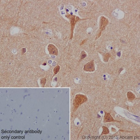
Immunohistochemical analysis of paraffin-embedded Human cerebral cortex tissue labeling SGTB with ab202419 at 1/100 dilution, followed by Goat Anti-Rabbit IgG H&L (HRP) (ab97051) secondary antibody at 1/500 dilution. Cytoplasmic and nuclear staining on Human cerebral cortex tissue is observed. Counter stained with Hematoxylin.Secondary antibody only control: Used PBS instead of primary antibody, secondary antibody is Goat Anti-Rabbit IgG H&L (HRP) (ab97051) at 1/500 dilution.
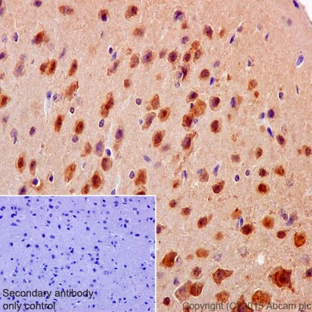
Immunohistochemical analysis of paraffin-embedded mouse cerebral cortex tissue labeling SGTB with ab202419 at 1/100 dilution, followed by Goat Anti-Rabbit IgG H&L (HRP) (ab97051) secondary antibody at 1/500 dilution. Cytoplasmic and nuclear staining on mouse cerebral cortex tissue is observed. Counter stained with Hematoxylin.Secondary antibody only control: Used PBS instead of primary antibody, secondary antibody is Goat Anti-Rabbit IgG H&L (HRP) (ab97051) at 1/500 dilution.
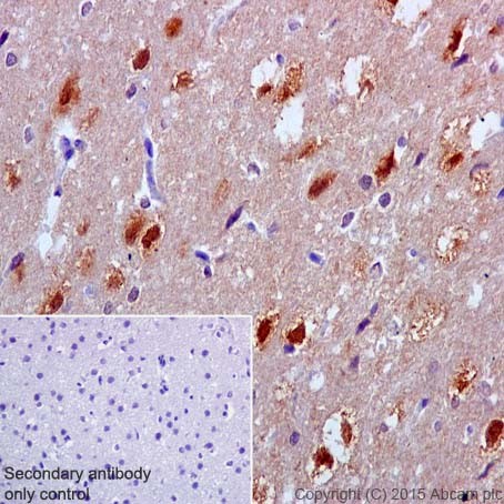
Immunohistochemical analysis of paraffin-embedded Rat cerebral cortex tissue labeling SGTB with ab202419 at 1/100 dilution, followed by Goat Anti-Rabbit IgG H&L (HRP) (ab97051) secondary antibody at 1/500 dilution. Cytoplasmic and nuclear staining on rat cerebral cortex tissue is observed. Counter stained with Hematoxylin.Secondary antibody only control: Used PBS instead of primary antibody, secondary antibody is Goat Anti-Rabbit IgG H&L (HRP) (ab97051) at 1/500 dilution.
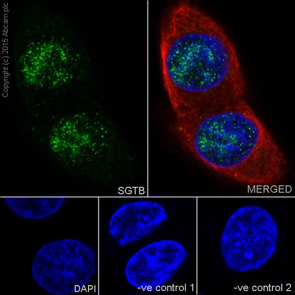
Immunofluorescent analysis of 4% paraformaldehyde-fixed, 0.1% Triton X-100 permeabilized SH-SY5Y (Human neuroblastoma from bone marrow cells) cells labeling SGTB with ab202419 at 1/100 dilution, followed by Goat anti-rabbit IgG (Alexa Fluor® 488) (ab150077) secondary antibody at 1/500 dilution (green). Confocal image showing nuclear and weakly cytoplasmic staining on SH-SY5Y cell line. The nuclear counter stain is DAPI (blue). Tubulin is detected with ab7291 (anti-Tubulin mouse mAb) at 1/1000 dilution and ab150120 (AlexaFluor®594 Goat anti-Mouse secondary) at 1/500 dilution (red).The negative controls are as follows:-ve control 1: ab202419 at 1/100 dilution followed by ab150120 (AlexaFluor®594 Goat anti-Mouse secondary) at 1/500 dilution.-ve control 2: ab7291 (anti-Tubulin mouse mAb) at 1/1000 dilution followed by ab150077 (Alexa Fluor®488 Goat Anti-Rabbit IgG H&L) at 1/500 dilution.
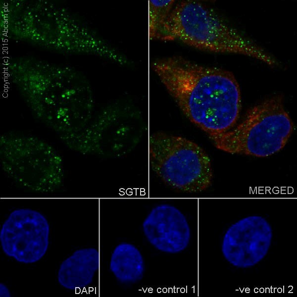
Immunofluorescent analysis of 4% paraformaldehyde-fixed, 0.1% Triton X-100 permeabilized U-87 MG (Human glioblastoma-astrocytoma epithelial cell line) cells labeling SGTB with ab202419 at 1/100 dilution, followed by Goat anti-rabbit IgG (Alexa Fluor® 488) (ab150077) secondary antibody at 1/500 dilution (green). Confocal image showing nuclear and cytoplasmic staining on U-87 MG cell line. The nuclear counter stain is DAPI (blue). Tubulin is detected with ab7291 (anti-Tubulin mouse mAb) at 1/1000 dilution and ab150120 (AlexaFluor®594 Goat anti-Mouse secondary) at 1/500 dilution (red).The negative controls are as follows:-ve control 1: ab202419 at 1/100 dilution followed by ab150120 (AlexaFluor®594 Goat anti-Mouse secondary) at 1/500 dilution.-ve control 2: ab7291 (anti-Tubulin mouse mAb) at 1/1000 dilution followed by ab150077 (Alexa Fluor®488 Goat Anti-Rabbit IgG H&L) at 1/500 dilution.
SGTB was immunoprecipitated from 1mg of Mouse brain whole cell lysate with ab202419 at 1/40 dilution. Western blot was performed from the immunoprecipitate using ab202419 at 1/1000 dilution. Anti-Rabbit IgG (HRP), specific to the non-reduced form of IgG, was used as secondary antibody at 1/1500 dilution.Lane 1: Mouse brain whole cell lysate 10 µg (Input). Lane 2: ab202419 IP in Mouse brain whole cell lysate. Lane 3: Rabbit monoclonal IgG (ab172730) instead of ab202419 in Mouse brain whole cell lysate.Blocking and dilution buffer and concentration: 5% NFDM/TBST.Exposure time: 1 second.