Anti-SENP2 antibody
| Name | Anti-SENP2 antibody |
|---|---|
| Supplier | Abcam |
| Catalog | ab58418 |
| Prices | $370.00 |
| Sizes | 100 µg |
| Host | Rabbit |
| Clonality | Polyclonal |
| Isotype | IgG |
| Applications | WB ELISA ICC/IF ICC/IF |
| Species Reactivities | Mouse, Rat, Human |
| Antigen | Synthetic peptide derived from an internal region of Human SENP2 |
| Description | Rabbit Polyclonal |
| Gene | SENP2 |
| Conjugate | Unconjugated |
| Supplier Page | Shop |
Product images
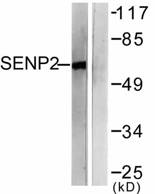 All lanes : Anti-SENP2 antibody (ab58418) at 1/500 dilutionLane 1 : MDA-MB-435 cell extractLane 2 : MDA-MB-435 cell extract with immunizing peptide
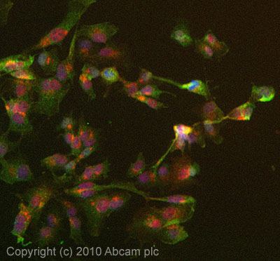 ICC/IF image of ab58418 stained HepG2 cells. The cells were 4% formaldehyde fixed (10 min) and then incubated in 1%BSA / 10% normal goat serum / 0.3M glycine in 0.1% PBS-Tween for 1h to permeabilise the cells and block non-specific protein-protein interactions. The cells were then incubated with the antibody (ab58418, 5µg/ml) overnight at +4°C. The secondary antibody (green) was Alexa Fluor® 488 goat anti-mouse IgG (H+L) used at a 1/1000 dilution for 1h. Alexa Fluor® 594 WGA was used to label plasma membranes (red) at a 1/200 dilution for 1h. DAPI was used to stain the cell nuclei (blue) at a concentration of 1.43µM.