![Overlay histogram showing HeLa cells stained with ab110201 (red line). The cells were fixed with 80% methanol (5 min)/ and then permeabilized with 0.1% PBS-Tween for 20 min. The cells were then incubated in 1x PBS / 10% normal goat serum / 0.3M glycine to block non-specific protein-protein interactions followed by the antibody (ab110201, 0.1μg/1x106 cells) for 30 min at 22°C. The secondary antibody used was Alexa Fluor® 488 goat anti-mouse IgG (H&L) (ab150113) at 1/2000 dilution for 30 min at 22°C. Isotype control antibody (black line) was mouse IgG1 [ICIGG1] (ab91353, 1μg/1x106 cells) used under the same conditions. Unlabelled sample (blue line) was also used as a control. Acquisition of >5,000 events were collected using a 20mW Argon ion laser (488nm) and 525/30 bandpass filter.](http://www.bioprodhub.com/system/product_images/ab_products/2/sub_4/25237_ab110201-7-ab110201FC.jpg)
Overlay histogram showing HeLa cells stained with ab110201 (red line). The cells were fixed with 80% methanol (5 min)/ and then permeabilized with 0.1% PBS-Tween for 20 min. The cells were then incubated in 1x PBS / 10% normal goat serum / 0.3M glycine to block non-specific protein-protein interactions followed by the antibody (ab110201, 0.1μg/1x106 cells) for 30 min at 22°C. The secondary antibody used was Alexa Fluor® 488 goat anti-mouse IgG (H&L) (ab150113) at 1/2000 dilution for 30 min at 22°C. Isotype control antibody (black line) was mouse IgG1 [ICIGG1] (ab91353, 1μg/1x106 cells) used under the same conditions. Unlabelled sample (blue line) was also used as a control. Acquisition of >5,000 events were collected using a 20mW Argon ion laser (488nm) and 525/30 bandpass filter.
![Anti-RPS7 antibody [3G4] (ab110201) + HeLa cells](http://www.bioprodhub.com/system/product_images/ab_products/2/sub_4/25238_RPS7-Primary-antibodies-ab110201-1.jpg)
Anti-RPS7 antibody [3G4] (ab110201) + HeLa cells
![Anti-RPS7 antibody [3G4] (ab110201) + NIH/3T3 cells](http://www.bioprodhub.com/system/product_images/ab_products/2/sub_4/25239_RPS7-Primary-antibodies-ab110201-2.jpg)
Anti-RPS7 antibody [3G4] (ab110201) + NIH/3T3 cells
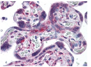
ab110201, at 5 µg/ml, staining RPS7 in Formalin-Fixed, Paraffin-Embedded Human placenta tissue.
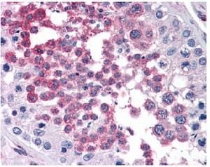
ab110201, at 5 µg/ml, staining RPS7 in Formalin-Fixed, Paraffin-Embedded Human testis tissue.
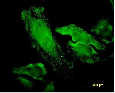
ab110201, at 20 µg/ml, staining RPS7 in HeLa cells.
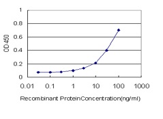
Detection limit for ab110201 is approximately 1 ng/ml as a capture antibody.