Anti-RPS3A antibody - C-terminal
| Name | Anti-RPS3A antibody - C-terminal |
|---|---|
| Supplier | Abcam |
| Catalog | ab174894 |
| Prices | $370.00 |
| Sizes | 400 µl |
| Host | Rabbit |
| Clonality | Polyclonal |
| Isotype | IgG |
| Applications | IHC-P WB FC |
| Species Reactivities | Human, Mouse, Rat, Bovine, Xenopus, Monkey |
| Antigen | Synthetic peptide within Human RPS3A aa 235-264 (C terminal) conjugated to Keyhole Limpet Haemocyanin (KLH) |
| Description | Rabbit Polyclonal |
| Gene | RPS3A |
| Conjugate | Unconjugated |
| Supplier Page | Shop |
Product images
 All lanes : Anti-RPS3A antibody - C-terminal (ab174894) at 1/100 dilutionLane 1 : Hela cell lysateLane 2 : A2058 cell lysateLysates/proteins at 15 µg per lane.
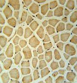 Immunohistochemistry analysis in formalin fixed, paraffin embedded Human skeletal muscle labeling RPS3A with ab174894 at 1/50 dilution, followed by peroxidase conjugation of the secondary antibody and DAB staining.
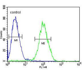 Flow cytometric analysis of Jurkat cells (right histogram) labeling RPS3A with ab174894 at 1/10 dilution, compared to a negative control cell (left histogram). FITC-conjugated goat-anti-rabbit secondary antibodies were used for the analysis.