![All lanes : Anti-RPS24 antibody [EPR16017(B)] (ab196652) at 1/5000 dilutionLane 1 : Jurkat (Human T cell leukemia cells from peripheral blood) whole cell lysateLane 2 : 293T (Human epithelial cells from embryonic kidney) whole cell lysateLysates/proteins at 10 µg per lane.SecondaryGoat Anti-Rabbit IgG, (H+L), Peroxidase conjugated at 1/1000 dilution](http://www.bioprodhub.com/system/product_images/ab_products/2/sub_4/25096_ab196652-236688-ab196652WBa.jpg)
All lanes : Anti-RPS24 antibody [EPR16017(B)] (ab196652) at 1/5000 dilutionLane 1 : Jurkat (Human T cell leukemia cells from peripheral blood) whole cell lysateLane 2 : 293T (Human epithelial cells from embryonic kidney) whole cell lysateLysates/proteins at 10 µg per lane.SecondaryGoat Anti-Rabbit IgG, (H+L), Peroxidase conjugated at 1/1000 dilution
![All lanes : Anti-RPS24 antibody [EPR16017(B)] (ab196652) at 1/1000 dilutionLane 1 : Mouse colon lysateLane 2 : Rat colon lysateLane 3 : Mouse liver lysateLysates/proteins at 10 µg per lane.SecondaryGoat Anti-Rabbit IgG, (H+L), Peroxidase conjugated at 1/1000 dilution](http://www.bioprodhub.com/system/product_images/ab_products/2/sub_4/25097_ab196652-236687-ab196652WBb.jpg)
All lanes : Anti-RPS24 antibody [EPR16017(B)] (ab196652) at 1/1000 dilutionLane 1 : Mouse colon lysateLane 2 : Rat colon lysateLane 3 : Mouse liver lysateLysates/proteins at 10 µg per lane.SecondaryGoat Anti-Rabbit IgG, (H+L), Peroxidase conjugated at 1/1000 dilution
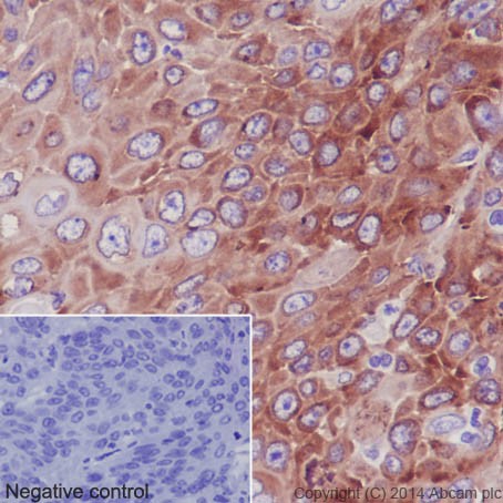
Immunohistochemical analysis of paraffin-embedded Human squamous cell carcinoma of cervix tissue labeling RPS24 with ab196652 at 1/100 dilution followed by Goat Anti-Rabbit IgG H&L (HRP) (ab97051) secondary antibody at 1/500 dilution. Cytoplasm staining on Human squamous cell carcinoma of cervix tissue is observed. Counter stained with Hematoxylin.
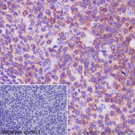
Immunohistochemical analysis of paraffin-embedded Human tonsil tissue labeling RPS24 with ab196652 at 1/100 dilution followed by Goat Anti-Rabbit IgG H&L (HRP) (ab97051) secondary antibody at 1/500 dilution. Cytoplasm staining on Human tonsil tissue is observed. Counter stained with Hematoxylin.
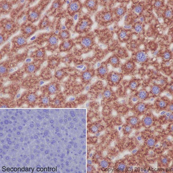
Immunohistochemical analysis of paraffin-embedded mouse liver tissue labeling RPS24 with ab196652 at 1/100 dilution followed by Goat Anti-Rabbit IgG H&L (HRP) (ab97051) secondary antibody at 1/500 dilution. Cytoplasm staining on mouse liver tissue is observed. Counter stained with Hematoxylin.
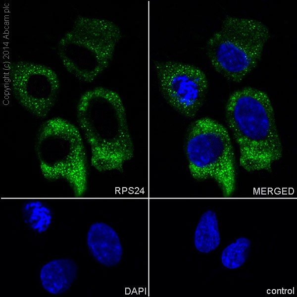
Immunofluorescent analysis of 4% paraformaldehyde-fixed, 0.1% Triton X-100 permeabilized SH-SY5Y (Human neuroblastoma from bone marrow cells) cells labeling RPS24 with ab196652 at 1/250 dilution, followed by AlexaFluor®488 Goat anti-Rabbit (ab150077) secondary antibody at 1/400 dilution (green). Cytoplasm staining on SH-SY5Y cell line is observed. The nuclear counter stain is DAPI (blue).