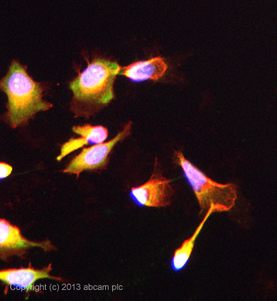
ab102986 stained HeLa cells. The cells were 100% methanol fixed (5 min) and then incubated in 1%BSA / 10% normal goat serum / 0.3M glycine in 0.1% PBS-Tween for 1h to permeabilise the cells and block non-specific protein-protein interactions. The cells were then incubated with the antibody ab102986 at 5µg/ml overnight at +4°C. The secondary antibody (green) was DyLight® 488 goat anti- rabbit (ab96899) IgG (H+L) used at a 1/250 dilution for 1h. Alexa Fluor® 594 WGA was used to label plasma membranes (red) at a 1/200 dilution for 1h. DAPI was used to stain the cell nuclei (blue) at a concentration of 1.43µM. This antibody also gave a positive result in formaldehyde fixed (4%, 10min) HeLa cells at 5ug/ml.
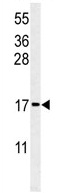
Anti-RPS24 antibody (ab102986) at 1/100 dilution + MCF7 cell line lysate at 35 µg
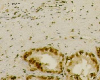
IHC-P image of RPS24 antibody (ab102986) on Seminal Vesicles. The sections were fixed in Paraformaldehyde and underwent heat mediated antigen retrieval using Novacastra (pH6). The sections were then blocked in 0.3% H2O2 solution for 10 minutes at 22°C.See Abreview