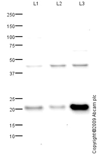
All lanes : Anti-RNase H2 subunit C antibody (ab89726) at 1 µg/mlLane 1 : HEK293 (Human embryonic kidney cell line) Whole Cell Lysate Lane 2 : HeLa (Human epithelial carcinoma cell line) Whole Cell LysateLane 3 : HepG2 (Human hepatocellular liver carcinoma cell line) Whole Cell LysateLysates/proteins at 10 µg per lane.SecondaryGoat polyclonal to Rabbit IgG - H&L - Pre-Adsorbed (HRP) at 1/3000 dilutiondeveloped using the ECL techniquePerformed under reducing conditions.
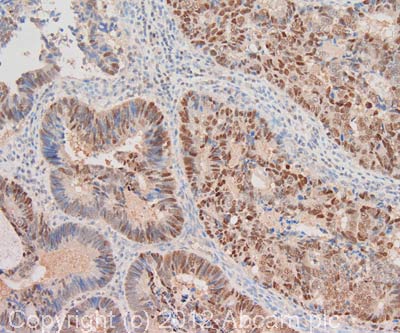
IHC image of RNase H2 subunit C staining in human colon adenocarcinoma formalin fixed paraffin embedded tissue section, performed on a Leica BondTM system using the standard protocol F. The section was pre-treated using heat mediated antigen retrieval with sodium citrate buffer (pH6, epitope retrieval solution 1) for 20 mins. The section was then incubated with ab89726, 1µg/ml, for 15 mins at room temperature and detected using an HRP conjugated compact polymer system. DAB was used as the chromogen. The section was then counterstained with haematoxylin and mounted with DPX. For other IHC staining systems (automated and non-automated) customers should optimize variable parameters such as antigen retrieval conditions, primary antibody concentration and antibody incubation times.
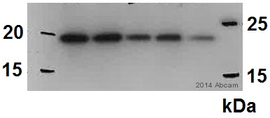
All lanes : Anti-RNase H2 subunit C antibody (ab89726) at 1 µg/mlLane 1 : Mouse Pei1 whole cell lysateLane 2 : Mouse Pei1 whole cell lysateLane 3 : Mouse Pei1 whole cell lysateLane 4 : Mouse Pei1 whole cell lysateLane 5 : Mouse Pei1 whole cell lysateLysates/proteins at 20 µg per lane.SecondaryHRP-conjugated goat anti-rabbit IgG polyclonal at 1/10000 dilutiondeveloped using the ECL techniquePerformed under reducing conditions.