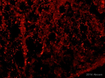
ab125275 staining RhoA (phospho S188) in mouse spinal cord tissue sections by Immunohistochemistry (IHC-Fr - frozen sections). Tissue was fixed with formaldehyde, permeabilized with citrate buffer and blocked with 5% serum for 1 hour at 25°C. Samples were incubated with primary antibody (5µg/ml) for 16 hours at 4°C. An Alexa Fluor® 568-conjugated goat anti-rabbit IgG polyclonal (1/200) was used as the secondary antibody.See Abreview
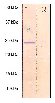
All lanes : Anti-RhoA (phospho S188) antibody (ab125275) at 1/500 dilutionLane 1 : MCF7 cell lysateLane 2 : MCF7 cell lysate with immunizing peptide
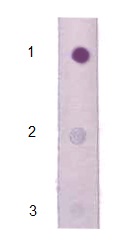
ab125275, at 1/2000 dilution, staining RhoA in 1 µg peptide blotted onto NC membrane (1) compared with non-phosphopeptide RhoA (2) and a non-related phosphopeptide (3) by Dot Blot.