![All lanes : Anti-Rho A + B + C antibody [EPR18299] (ab188103) at 1/20000 dilutionLane 1 : Human RhoA full length proteinLane 2 : Human RhoB full length proteinLysates/proteins at 0.01 µg per lane.SecondaryGoat Anti-Rabbit IgG, (H+L),Peroxidase conjugated at 1/1000 dilution](http://www.bioprodhub.com/system/product_images/ab_products/2/sub_4/22789_ab188103-242941-188103.jpg)
All lanes : Anti-Rho A + B + C antibody [EPR18299] (ab188103) at 1/20000 dilutionLane 1 : Human RhoA full length proteinLane 2 : Human RhoB full length proteinLysates/proteins at 0.01 µg per lane.SecondaryGoat Anti-Rabbit IgG, (H+L),Peroxidase conjugated at 1/1000 dilution
![Anti-Rho A + B + C antibody [EPR18299] (ab188103) at 1/500 dilution + Human RhoC full length protein at 0.01 µgSecondaryGoat Anti-Rabbit IgG, (H+L),Peroxidase conjugated at 1/1000 dilution](http://www.bioprodhub.com/system/product_images/ab_products/2/sub_4/22790_ab188103-242942-188103WBb.jpg)
Anti-Rho A + B + C antibody [EPR18299] (ab188103) at 1/500 dilution + Human RhoC full length protein at 0.01 µgSecondaryGoat Anti-Rabbit IgG, (H+L),Peroxidase conjugated at 1/1000 dilution
![All lanes : Anti-Rho A + B + C antibody [EPR18299] (ab188103) at 1/2000 dilutionLane 1 : HepG2 (Human liver hepatocellular carcinoma) whole cell lysateLane 2 : HeLa (Human epithelial cells from cervix adenocarcinoma) whole cell lysateLane 3 : Jurkat (Human T cell leukemia cells from peripheral blood) whole cell lysateLysates/proteins at 10 µg per lane.SecondaryGoat Anti-Rabbit IgG, (H+L),Peroxidase conjugated at 1/1000 dilution](http://www.bioprodhub.com/system/product_images/ab_products/2/sub_4/22791_ab188103-242943-1881033.jpg)
All lanes : Anti-Rho A + B + C antibody [EPR18299] (ab188103) at 1/2000 dilutionLane 1 : HepG2 (Human liver hepatocellular carcinoma) whole cell lysateLane 2 : HeLa (Human epithelial cells from cervix adenocarcinoma) whole cell lysateLane 3 : Jurkat (Human T cell leukemia cells from peripheral blood) whole cell lysateLysates/proteins at 10 µg per lane.SecondaryGoat Anti-Rabbit IgG, (H+L),Peroxidase conjugated at 1/1000 dilution
![All lanes : Anti-Rho A + B + C antibody [EPR18299] (ab188103) at 1/2000 dilutionLane 1 : Human fetal brain lysateLane 2 : Human fetal kidney lysateLysates/proteins at 10 µg per lane.SecondaryAnti-Rabbit IgG (HRP), specific to the non-reduced form of IgG at 1/1000 dilution](http://www.bioprodhub.com/system/product_images/ab_products/2/sub_4/22792_ab188103-242946-1881034.jpg)
All lanes : Anti-Rho A + B + C antibody [EPR18299] (ab188103) at 1/2000 dilutionLane 1 : Human fetal brain lysateLane 2 : Human fetal kidney lysateLysates/proteins at 10 µg per lane.SecondaryAnti-Rabbit IgG (HRP), specific to the non-reduced form of IgG at 1/1000 dilution
![All lanes : Anti-Rho A + B + C antibody [EPR18299] (ab188103) at 1/2000 dilutionLane 1 : Mouse brain lysateLane 2 : Mouse kidney lysateLane 3 : Rat brain lysateLane 4 : Rat kidney lysateLane 5 : C6 (Rat glial tumor cells) whole cell lysateLane 6 : RAW 264.7 (Mouse macrophage cells transformed with Abelson murine leukemia virus) whole cell lysateLane 7 : PC-12 (Rat adrenal gland pheochromocytoma) whole cell lysateLane 8 : NIH/3T3 (Mouse embyro fibroblast cells) whole cell lysateLysates/proteins at 10 µg per lane.SecondaryGoat Anti-Rabbit IgG, (H+L),Peroxidase conjugated at 1/1000 dilution](http://www.bioprodhub.com/system/product_images/ab_products/2/sub_4/22793_ab188103-242947-1881035.jpg)
All lanes : Anti-Rho A + B + C antibody [EPR18299] (ab188103) at 1/2000 dilutionLane 1 : Mouse brain lysateLane 2 : Mouse kidney lysateLane 3 : Rat brain lysateLane 4 : Rat kidney lysateLane 5 : C6 (Rat glial tumor cells) whole cell lysateLane 6 : RAW 264.7 (Mouse macrophage cells transformed with Abelson murine leukemia virus) whole cell lysateLane 7 : PC-12 (Rat adrenal gland pheochromocytoma) whole cell lysateLane 8 : NIH/3T3 (Mouse embyro fibroblast cells) whole cell lysateLysates/proteins at 10 µg per lane.SecondaryGoat Anti-Rabbit IgG, (H+L),Peroxidase conjugated at 1/1000 dilution
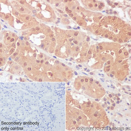
Immunohistochemical analysis of paraffin-embedded Human stomach tissue labeling Rho A + B + C with ab188103 at 1/4000 dilution, followed by Goat Anti-Rabbit IgG H&L (HRP) (ab97051) secondary antibody at 1/500 dilution. Cytoplasmic and nuclear staining on epithelial cells of Human stomach is observed. Counter stained with Hematoxylin.Secondary antibody only control: Used PBS instead of primary antibody, secondary antibody is Goat Anti-Rabbit IgG H&L (HRP) (ab97051) at 1/500 dilution.
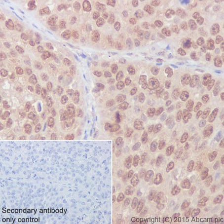
Immunohistochemical analysis of paraffin-embedded Human squamous cell carcinoma of lung tissue labeling Rho A + B + C with ab188103 at 1/4000 dilution, followed by Goat Anti-Rabbit IgG H&L (HRP) (ab97051) secondary antibody at 1/500 dilution. Cytoplasmic and nuclear staining on cancer cells of squamous cell carcinoma of lung is observed. Counter stained with Hematoxylin.Secondary antibody only control: Used PBS instead of primary antibody, secondary antibody is Goat Anti-Rabbit IgG H&L (HRP) (ab97051) at 1/500 dilution.
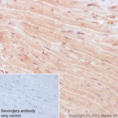
Immunohistochemical analysis of paraffin-embedded Mouse muscle tissue labeling Rho A + B + C with ab188103 at 1/4000 dilution, followed by Goat Anti-Rabbit IgG H&L (HRP) (ab97051) secondary antibody at 1/500 dilution. Cytoplasmic and nuclear staining on mouse muscle is observed. Counter stained with Hematoxylin.Secondary antibody only control: Used PBS instead of primary antibody, secondary antibody is Goat Anti-Rabbit IgG H&L (HRP) (ab97051) at 1/500 dilution.
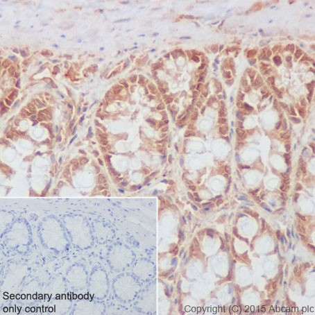
Immunohistochemical analysis of paraffin-embedded Rat colon tissue labeling Rho A + B + C with ab188103 at 1/4000 dilution, followed by Goat Anti-Rabbit IgG H&L (HRP) (ab97051) secondary antibody at 1/500 dilution. Cytoplasmic and nuclear staining on epithelial cells of Rat colon is observed. Counter stained with Hematoxylin.Secondary antibody only control: Used PBS instead of primary antibody, secondary antibody is Goat Anti-Rabbit IgG H&L (HRP) (ab97051) at 1/500 dilution.
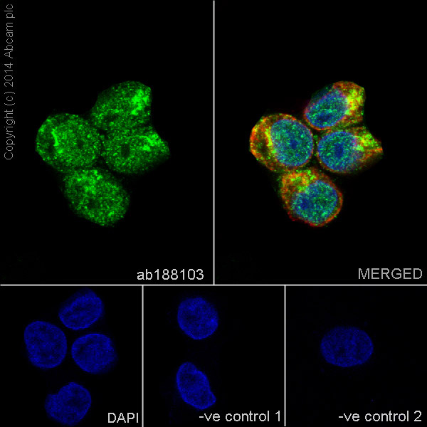
Immunofluorescent analysis of 4% paraformaldehyde-fixed, 0.1% Triton X-100 permeabilized HepG2 (Human liver hepatocellular carcinoma) cells labeling Rho A + B + C with ab188103 at 1/500 dilution, followed by Goat anti-rabbit IgG (Alexa Fluor® 488) (ab150077) secondary antibody at 1/500 dilution (green). Confocal image showing both nuclear and cytoplasmic staining on HepG2 cells. The nuclear counter stain is DAPI (blue). Tubulin is detected with ab7291 (anti-Tubulin mouse mAb) at 1/1000 dilution and ab150120 (AlexaFluor®594 Goat anti-Mouse secondary) at 1/500 dilution (red).The negative controls are as follows;-ve control 1: ab188103 at 1/500 dilution followed by ab150120 (AlexaFluor®594 Goat anti-Mouse secondary) at 1/500 dilution.-ve control 2: ab7291 (anti-Tubulin mouse mAb) at 1/1000 dilution followed by ab150077 (Alexa Fluor®488 Goat Anti-Rabbit IgG H&L) at 1/500 dilution.
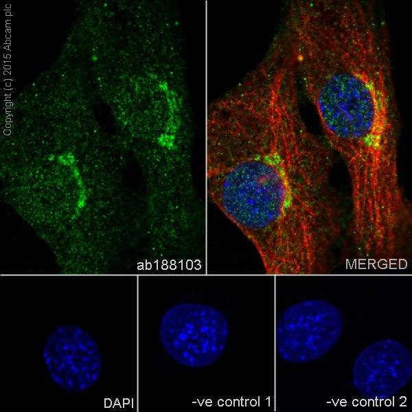
Immunofluorescent analysis of 4% paraformaldehyde-fixed, 0.1% Triton X-100 permeabilized NIH/3T3 (Mouse embyro fibroblast cells) cells labeling to RhoA + RhoB + RhoC with ab188103 at 1/500 dilution, followed by Goat anti-rabbit IgG (Alexa Fluor® 488) (ab150077) secondary antibody at 1/500 dilution (green). Confocal image showing both nuclear and cytoplasmic staining on NIH/3T3 cells. The nuclear counter stain is DAPI (blue). Tubulin is detected with ab7291 (anti-Tubulin mouse mAb) at 1/1000 dilution and ab150120 (AlexaFluor®594 Goat anti-Mouse secondary) at 1/500 dilution (red).The negative controls are as follows;-ve control 1: ab188103 at 1/500 dilution followed by ab150120 (AlexaFluor®594 Goat anti-Mouse secondary) at 1/500 dilution.-ve control 2: ab7291 (anti-Tubulin mouse mAb) at 1/1000 dilution followed by ab150077 (Alexa Fluor®488 Goat Anti-Rabbit IgG H&L) at 1/500 dilution.
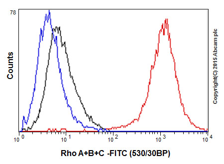
Flow cytometric analysis of 2% paraformaldehyde-fixed Jurkat (Human T cell leukemia cells from peripheral blood) cells labeling Rho A + B + C with ab188103 at 1/300 dilution (red) compared with a rabbit monoclonal IgG isotype control (black) and an unlabelled control (cells without incubation with primary antibody and secondary antibody; blue). Goat anti rabbit IgG (FITC) at 1/150 dilution was used as the secondary antibody.