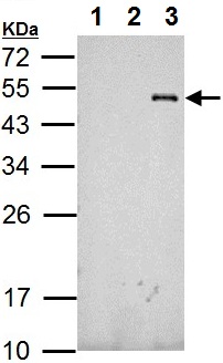
All lanes : Anti-RFP antibody (ab152123) at 1/10000 dilutionLane 1 : Non-transfected 293T whole cell lysateLane 2 : GFP-transfected 293T whole cell lysateLane 3 : RFP-transfected 293T whole cell lysateLysates/proteins at 5 µg per lane.
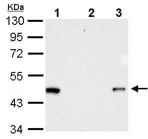
Immunoprecipitation of RFP-tagged protein. HeLa transfected lysates were subjected to immunoprecipitation using: Lane 1: 15 µg of HeLa transfected lysate Lane 2: normal rabbit IgG Lane 3: 2.5 µg ab152123 Detection ab152123 at 1/10000 dilution
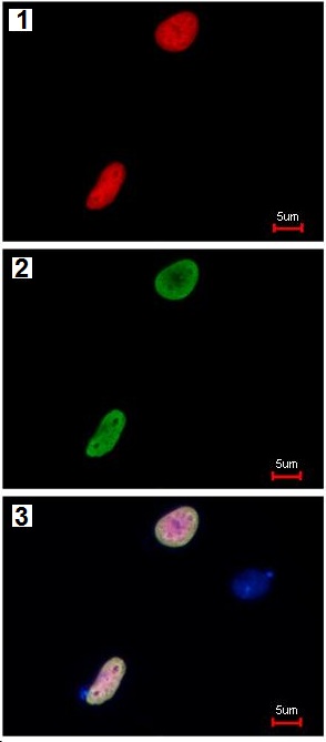
Immunofluorescence analysis of RFP-transfected HeLa cells labeling RFP with ab152123: (1) RFP is expressed in the tranfected cell, (2) The cell expressing RFP is detected using ab152123 at 1/5000 followed by Alexa Fluor 488 at 1/500 Goat anti-Rabbit IgG, (3) Merged with DNA probe, the lower cell represents a negtive control.