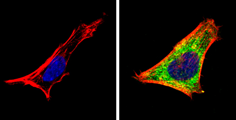
Immunocytochemistry/Immunofluorescence analysis of SH-SY5Y cells labeling Retinoic Acid Receptor (green) with ab5423 at 1/100. F-Actin staining with Phalloidin (red) and nuclei with DAPI or Hoechst (blue). Cells were fixed with formalin, permeabilized with 0.1% Triton X-100 in TBS for 5-10 minutes and blocked with 3% BSA in PBS for 30 minutes at room temperature. Cells were incubated with the primary antibody in 3% BSA in PBS overnight at 4°C. A DyLight-conjugated secondary antibody was used. 60X magnification. Left - negative control.
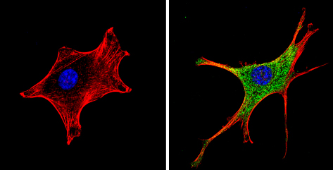
Immunocytochemistry/Immunofluorescence analysis of NIH-3T3 cells labeling Retinoic Acid Receptor (green) with ab5423 at 1/100. F-Actin staining with Phalloidin (red) and nuclei with DAPI or Hoechst (blue). Cells were fixed with formalin, permeabilized with 0.1% Triton X-100 in TBS for 5-10 minutes and blocked with 3% BSA in PBS for 30 minutes at room temperature. Cells were incubated with the primary antibody in 3% BSA in PBS overnight at 4°C. A DyLight-conjugated secondary antibody was used. 60X magnification. Left - negative control.
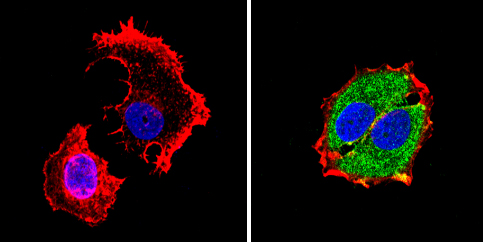
Immunocytochemistry/Immunofluorescence analysis of MCF-7 cells labeling Retinoic Acid Receptor (green) with ab5423 at 1/100. F-Actin staining with Phalloidin (red) and nuclei with DAPI or Hoechst (blue). Cells were fixed with formalin, permeabilized with 0.1% Triton X-100 in TBS for 5-10 minutes and blocked with 3% BSA in PBS for 30 minutes at room temperature. Cells were incubated with the primary antibody in 3% BSA in PBS overnight at 4°C. A DyLight-conjugated secondary antibody was used. 60X magnification. Left - negative control.