![All lanes : Anti-RCN1 antibody [EPR17163] - C-terminal (ab198996) at 1/10000 dilutionLane 1 : 293 (Human epithelial cells from embryonic kidney) cell lysateLane 2 : HeLa (Human epithelial cells from cervix adenocarcinoma) cell lysateLysates/proteins at 10 µg per lane.SecondaryGoat Anti-Rabbit IgG, (H+L), Peroxidase conjugated at 1/1000 dilutiondeveloped using the ECL technique](http://www.bioprodhub.com/system/product_images/ab_products/2/sub_4/21749_ab198996-241449-ab198996WB1.jpg)
All lanes : Anti-RCN1 antibody [EPR17163] - C-terminal (ab198996) at 1/10000 dilutionLane 1 : 293 (Human epithelial cells from embryonic kidney) cell lysateLane 2 : HeLa (Human epithelial cells from cervix adenocarcinoma) cell lysateLysates/proteins at 10 µg per lane.SecondaryGoat Anti-Rabbit IgG, (H+L), Peroxidase conjugated at 1/1000 dilutiondeveloped using the ECL technique
![All lanes : Anti-RCN1 antibody [EPR17163] - C-terminal (ab198996) at 1/1000 dilutionLane 1 : Raw264.7 (Mouse macrophage cells transformed with Abelson murine leukemia virus) cell lysateLane 2 : Rat heart tissue lysateLysates/proteins at 10 µg per lane.SecondaryGoat Anti-Rabbit IgG, (H+L), Peroxidase conjugated at 1/1000 dilutiondeveloped using the ECL technique](http://www.bioprodhub.com/system/product_images/ab_products/2/sub_4/21750_ab198996-241450-ab198996WB2.jpg)
All lanes : Anti-RCN1 antibody [EPR17163] - C-terminal (ab198996) at 1/1000 dilutionLane 1 : Raw264.7 (Mouse macrophage cells transformed with Abelson murine leukemia virus) cell lysateLane 2 : Rat heart tissue lysateLysates/proteins at 10 µg per lane.SecondaryGoat Anti-Rabbit IgG, (H+L), Peroxidase conjugated at 1/1000 dilutiondeveloped using the ECL technique
![Immunohistochemical analysis of paraffin-embedded Human cerebral cortex tissue labeling RCN1 with ab198996 at 1/1600, followed by Goat Anti-Rabbit IgG H&L (HRP) (ab97051) at 1/500. Cytoplasm staining on Human cerebral cortex tissue is observed. Subcellular location Endoplasmic reticulum lumen [UniProt]. Counter stained with Hematoxylin.Negative control: Used PBS instead of primary antibody, secondary antibody is Goat Anti-Rabbit IgG H&L (HRP) (ab97051) at 1/500 dilution.](http://www.bioprodhub.com/system/product_images/ab_products/2/sub_4/21751_ab198996-241451-ab198996IHC1.jpg)
Immunohistochemical analysis of paraffin-embedded Human cerebral cortex tissue labeling RCN1 with ab198996 at 1/1600, followed by Goat Anti-Rabbit IgG H&L (HRP) (ab97051) at 1/500. Cytoplasm staining on Human cerebral cortex tissue is observed. Subcellular location Endoplasmic reticulum lumen [UniProt]. Counter stained with Hematoxylin.Negative control: Used PBS instead of primary antibody, secondary antibody is Goat Anti-Rabbit IgG H&L (HRP) (ab97051) at 1/500 dilution.
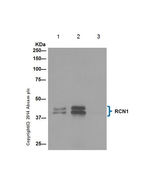
RCN1 was immunoprecipitated from 1mg of HeLa (Human epithelial cells from cervix adenocarcinoma) whole cell extract with ab198996 at 1/100. Western blot was performed from the immunoprecipitate using ab198996 at 1/1000. Anti-Rabbit IgG (HRP), specific to the non-reduced form of IgG, was used as secondary antibody at 1/1500.Lane 1: HeLa (Human epithelial cells from cervix adenocarcinoma) whole cell extract. 10ug (Input.Lane 2: HeLa whole cell extract.Lane 3: Rabbit monoclonal IgG (ab172730) instead of ab198996 in HeLa whole cell extract.The expression profile observed is consistent with what has been described in the literature PMID: 8416973. Blocking and dilution buffer and concentration: 5% NFDM/TBST.