![All lanes : Anti-RASEF antibody [EPR16349] (ab194827) at 1/10000 dilutionLane 1 : Mouse brain lysateLane 2 : Mouse heart lysateLane 3 : Mouse spleen lysateLane 4 : Rat brain lysateLane 5 : Rat heart lysateLane 6 : C6 cell lysateLane 7 : RAW 264.7 cell lysateLane 8 : PC12 cell lysateLane 9 : NIH 3T3 cell lysateLysates/proteins at 10 µg per lane.SecondaryGoat Anti-Rabbit IgG, (H+L), Peroxidase conjugated at 1/1000 dilution](http://www.bioprodhub.com/system/product_images/ab_products/2/sub_4/20892_ab194827-232559-ab194827-wb-3.jpg)
All lanes : Anti-RASEF antibody [EPR16349] (ab194827) at 1/10000 dilutionLane 1 : Mouse brain lysateLane 2 : Mouse heart lysateLane 3 : Mouse spleen lysateLane 4 : Rat brain lysateLane 5 : Rat heart lysateLane 6 : C6 cell lysateLane 7 : RAW 264.7 cell lysateLane 8 : PC12 cell lysateLane 9 : NIH 3T3 cell lysateLysates/proteins at 10 µg per lane.SecondaryGoat Anti-Rabbit IgG, (H+L), Peroxidase conjugated at 1/1000 dilution

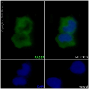
Immunofluorescent analysis of HeLa cells labeling RASEF with ab194827 at 1/50 dilution. A Goat anti rabbit IgG (Alexa Fluor488) at 1/400 dilution (ab150077) was used as secondary antibody. Cells were fixed with 4% paraformaldehyde and permeabilized with 0.1% triton X-100. Counterstain: DAPI. Cytoplasm staining on HeLa cell line was observed.
![Anti-RASEF antibody [EPR16349] (ab194827) at 1/10000 dilution + K562 cell lysate at 20 µgSecondaryGoat Anti-Rabbit IgG, (H+L), Peroxidase conjugated at 1/1000 dilution](http://www.bioprodhub.com/system/product_images/ab_products/2/sub_4/20894_ab194827-232558-ab194827-wb-2.jpg)
Anti-RASEF antibody [EPR16349] (ab194827) at 1/10000 dilution + K562 cell lysate at 20 µgSecondaryGoat Anti-Rabbit IgG, (H+L), Peroxidase conjugated at 1/1000 dilution
![All lanes : Anti-RASEF antibody [EPR16349] (ab194827) at 1/1000 dilutionLane 1 : A549 cell lysateLane 2 : A431 cell lysateLane 3 : Human testis lysateLysates/proteins at 20 µg per lane.SecondaryGoat Anti-Rabbit IgG, (H+L), Peroxidase conjugated at 1/1000 dilution](http://www.bioprodhub.com/system/product_images/ab_products/2/sub_4/20895_ab194827-232557-ab194827-wb-1.jpg)
All lanes : Anti-RASEF antibody [EPR16349] (ab194827) at 1/1000 dilutionLane 1 : A549 cell lysateLane 2 : A431 cell lysateLane 3 : Human testis lysateLysates/proteins at 20 µg per lane.SecondaryGoat Anti-Rabbit IgG, (H+L), Peroxidase conjugated at 1/1000 dilution

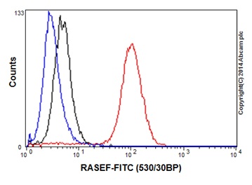
Flow cytometry analysis of K562 cells labeling RASEF using ab194827 at 1/50 dilution (Red). A Goat anti rabbit IgG (FITC) at 1/150 dilution was used as secondary antibody. Cells were fixed with 2% paraformaldehyde. Cells without incubation with primary antibody and secondary antibody: Blue. Rabbit monoclonal IgG was used as isotype control (Black).
![All lanes : Anti-RASEF antibody [EPR16349] (ab194827) at 1/10000 dilutionLane 1 : Mouse brain lysateLane 2 : Mouse heart lysateLane 3 : Mouse spleen lysateLane 4 : Rat brain lysateLane 5 : Rat heart lysateLane 6 : C6 cell lysateLane 7 : RAW 264.7 cell lysateLane 8 : PC12 cell lysateLane 9 : NIH 3T3 cell lysateLysates/proteins at 10 µg per lane.SecondaryGoat Anti-Rabbit IgG, (H+L), Peroxidase conjugated at 1/1000 dilution](http://www.bioprodhub.com/system/product_images/ab_products/2/sub_4/20892_ab194827-232559-ab194827-wb-3.jpg)

![Anti-RASEF antibody [EPR16349] (ab194827) at 1/10000 dilution + K562 cell lysate at 20 µgSecondaryGoat Anti-Rabbit IgG, (H+L), Peroxidase conjugated at 1/1000 dilution](http://www.bioprodhub.com/system/product_images/ab_products/2/sub_4/20894_ab194827-232558-ab194827-wb-2.jpg)
![All lanes : Anti-RASEF antibody [EPR16349] (ab194827) at 1/1000 dilutionLane 1 : A549 cell lysateLane 2 : A431 cell lysateLane 3 : Human testis lysateLysates/proteins at 20 µg per lane.SecondaryGoat Anti-Rabbit IgG, (H+L), Peroxidase conjugated at 1/1000 dilution](http://www.bioprodhub.com/system/product_images/ab_products/2/sub_4/20895_ab194827-232557-ab194827-wb-1.jpg)
