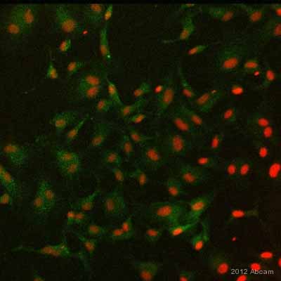
ab10590 staining RANTES in human endothelial cells by Immunocytochemistry/ Immunofluorescence.Cells were fixed in paraformaldehyde, permeabilized using Triton, blocked with 1% BSA for 1 hour at 25°C and then incubated with ab10590 at 2µg/ml for 1 hour at 25°C. The secondary used was an FITC conjugated goat anti-rabbit polyclonal used at a 1/100 dilution. Counter stained with TO-PRO3.See Abreview
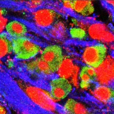
ab10590 staining RANTES in Human liver cancer metastases by Immunohistochemistry (Frozen sections). Tissue was fixed with paraformaldehyde, blocked with 5% serum for 1 hour at 24°C and permeabilized with Triton X-100. Samples were incubated with primary antibody (in PBS + 0.3% Triton X-100 + 1% BSA) for 3 hours at 24°C. An AlexaFluor®488-conjugated donkey anti-goat IgG polyclonal was used as the secondary antibody.See Abreview