CEA/CD66e (CB30) Mouse mAb
| Name | CEA/CD66e (CB30) Mouse mAb |
|---|---|
| Supplier | Cell Signaling Technology |
| Catalog | 2383 |
| Prices | $246.00 |
| Sizes | 100 µl (10 western blots) |
| Host | Mouse |
| Clonality | Monoclonal |
| Isotype | IgG1 |
| Clone | CB30 |
| Applications | WB IHC-P ICC/IF FC |
| Species Reactivities | Human |
| Antigen | Monoclonal antibody (Isotype: IgG1) is produced by immunizing mice with human CEA. |
| Description | Mouse Monoclonal |
| Gene | CEACAM5 |
| Supplier Page | Shop |
Product images
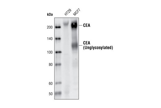 Western blot analysis of extracts from HT29 and MCF7 cells, using CEA/CD66e (CB30) Mouse mAb.
 Immunohistochemical analysis of paraffin-embedded human colon carcinoma, using CEA/CD66e (CB30) Mouse mAb.
 Confocal immunofluorescent analysis of MCF7 (+) and PANC-1 (-) cells using CEA/CD66e (CB30) Mouse mAb (green). Actin filaments were labeled with DY-554 Phalloidin (red). Blue pseudocolor = DRAQ5 ® #4084 (fluorescent DNA dye).
 Flow cytometric analysis of HT-29 cells, using CEA/CD66e (CB30) Mouse mAb (blue) compared to a nonspecific negative control antibody (red).