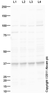
All lanes : Anti-RAD51AP1 antibody (ab101321) at 1 µg/mlLane 1 : HeLa (Human epithelial carcinoma cell line) Whole Cell LysateLane 2 : HEK293 (Human embryonic kidney cell line) Whole Cell LysateLane 3 : U2OS (Human osteosarcoma cell line) Whole Cell Lysate Lane 4 : Raji (Human Burkitt's lymphoma cell line) Whole Cell LysateLysates/proteins at 10 µg per lane.SecondaryGoat Anti-Rabbit IgG H&L (HRP) preadsorbed (ab97080) at 1/5000 dilutiondeveloped using the ECL techniquePerformed under reducing conditions.
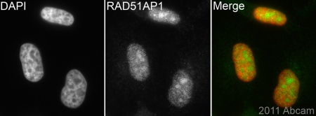
ab101321 (1/200) staining RAD51AP1 in HeLa cells (green). Cells were fixed in paraformaldehyde, permeabilized with 0.5% Triton X-100/ PBS and counterstained with DAPI in order to highlight the nucleus (red). For further experimental details please refer to Abreview.See Abreview