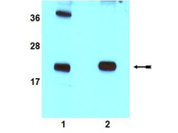
All lanes : Anti-Rac1 antibody (ab78139) at 1/1000 dilutionLane 1 : HeLa cell lysateLane 2 : MCF7 cell lysateSecondaryGoat anti-Rabbit HRP-conjugated at 1/5000 dilutionObserved band size : 22 kDa (why is the actual band size different from the predicted?)Additional bands at : 38 kDa. We are unsure as to the identity of these extra bands.
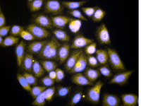
Immunofluorescence analysis of HeLa cells using ab78139 at 1/50 dilution (yellow) in conjunction with a Donkey anti-Rabbit IgG Cy3 conjugated. Hela cells were fixed with 3.7% paraformaldehyde and permeabilized with a non-ionic detergent. The cells are dual stained with DAPI (blue).
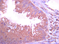
Immunohistochemistry analysis of paraffin-embedded Human tubular colorectal carcinoma using ab78139 at 1/1000 dilution with an HRP-DAB detection system. Tissue pretreated with citrate pH6.0 antigen retrieval. Immunoreactivity is seen primarily as diffusive plasma membrane staining.
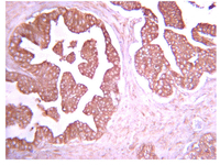
Immunohistochemistry analysis of paraffin-embedded Human prostate carcinoma using ab78139 at 1/1000 dilution with an HRP-DAB detection system. Tissue pretreated with citrate pH6.0 antigen retrieval. Immunoreactivity is seen primarily as diffusive plasma membrane staining with some stromal background staining.
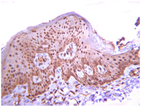
Immunohistochemistry analysis of paraffin-embedded Human squamous cells carcinoma using ab78139 at 1/1000 dilution with an HRP-DAB detection system. Tissue pretreated with citrate pH6.0 antigen retrieval. Immunoreactivity is seen primarily as diffusive nuclear staining pattern.