ab155953 showing +ve staining in Human thyroid gland carcinoma.
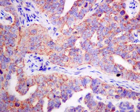
ab155953 showing +ve staining in Human ovarian carcinoma.
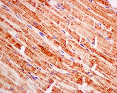
ab155953 showing +ve staining in Human heart.
ab155953 showing +ve staining in Human gastric adenocarcinoma.
![All lanes : Anti-Pyruvate Dehydrogenase E1 beta subunit antibody [EPR11096(B)] (ab155953) at 1/1000 dilutionLane 1 : HepG2 cell lysateLane 2 : Human fetal heart tissue lysateLane 3 : HeLa cell lysateLane 4 : A375 cell lysateLysates/proteins at 10 µg per lane.SecondaryGoat anti-rabbit HRP at 1/2000 dilution](http://www.bioprodhub.com/system/product_images/ab_products/2/sub_4/19023_Pyruvate-Dehydrogenase-E1-beta-subunit-Primary-antibodies-ab155953-1.JPG)
All lanes : Anti-Pyruvate Dehydrogenase E1 beta subunit antibody [EPR11096(B)] (ab155953) at 1/1000 dilutionLane 1 : HepG2 cell lysateLane 2 : Human fetal heart tissue lysateLane 3 : HeLa cell lysateLane 4 : A375 cell lysateLysates/proteins at 10 µg per lane.SecondaryGoat anti-rabbit HRP at 1/2000 dilution
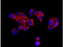
Immunofluorescent analysis of HepG2 cells, labeling Pyruvate Dehydrogenase E1 beta subunit with ab155953 at 1/100 dilution.
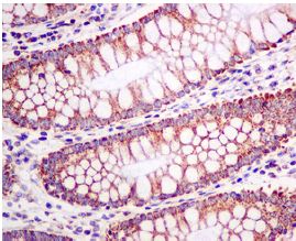
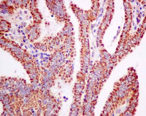
Immunohistochemical analysis of paraffin-embedded Human colon tissue, labeling Pyruvate Dehydrogenase E1 beta subunit with ab155953 at 1/50 dilution.
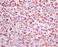
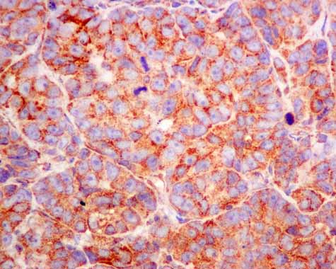
Immunohistochemical analysis of paraffin-embedded Human pancreas tissue, labeling Pyruvate Dehydrogenase E1 beta subunit with ab155953 at 1/50 dilution.
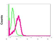
Flow cytometric analysis of permeabilized A375 cells labeling Pyruvate Dehydrogenase E1 beta subunit with ab155953 at 1/10 dilution (red) compared with a rabbit IgG negative control (green).