![All lanes : Anti-PSMA4 [EPR5831(2)] antibody (ab191403) at 1/10000 dilutionLane 1 : Jurkat cell lysateLane 2 : K562 cell lysateLane 3 : HeLa cell lysateLane 4 : JAR cell lysateLysates/proteins at 20 µg per lane.Secondarygoat anti-rabbit IgG, (H+L), peroxidase conjugated at 1/1000 dilutiondeveloped using the ECL technique](http://www.bioprodhub.com/system/product_images/ab_products/2/sub_4/17803_ab191403-228686-ab191403WB1.jpg)
All lanes : Anti-PSMA4 [EPR5831(2)] antibody (ab191403) at 1/10000 dilutionLane 1 : Jurkat cell lysateLane 2 : K562 cell lysateLane 3 : HeLa cell lysateLane 4 : JAR cell lysateLysates/proteins at 20 µg per lane.Secondarygoat anti-rabbit IgG, (H+L), peroxidase conjugated at 1/1000 dilutiondeveloped using the ECL technique
![All lanes : Anti-PSMA4 [EPR5831(2)] antibody (ab191403) at 1/1000 dilutionLane 1 : rat C6 cell lysateLane 2 : mouse Raw 264.7 cell lysateLane 3 : rat PC12 cell lysateLane 4 : mouse NIH 3T3 cell lysateLysates/proteins at 10 µg per lane.Secondarygoat anti-rabbit IgG, (H+L), peroxidase conjugated at 1/1000 dilutiondeveloped using the ECL technique](http://www.bioprodhub.com/system/product_images/ab_products/2/sub_4/17804_ab191403-228685-ab191403WB2.jpg)
All lanes : Anti-PSMA4 [EPR5831(2)] antibody (ab191403) at 1/1000 dilutionLane 1 : rat C6 cell lysateLane 2 : mouse Raw 264.7 cell lysateLane 3 : rat PC12 cell lysateLane 4 : mouse NIH 3T3 cell lysateLysates/proteins at 10 µg per lane.Secondarygoat anti-rabbit IgG, (H+L), peroxidase conjugated at 1/1000 dilutiondeveloped using the ECL technique
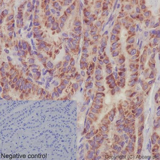
Immunohistochemical analysis of paraffin-embedded, Human papillary adenocarcinoma of thyroid tissue labeling PSMA4 with ab191403 at a 1/2000 dilution (1μg/ml). Counter stained with hematoxylin. In the negative control PBS was used instead of primary antibody.
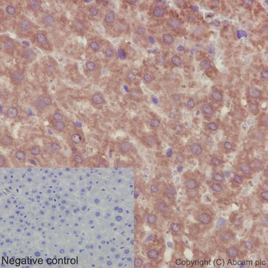
Immunohistochemical analysis of paraffin-embedded, mouse liver tissue labeling PSMA4 with ab191403 at a 1/2000 dilution (1μg/ml). Counter stained with hematoxylin. In the negative control PBS was used instead of primary antibody.
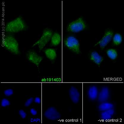
Immunofluorescence analysis of, paraformaldehyde-fixed, HeLa cells labeling PSMA4 with ab191403 at a 1/500 dilution ( 4 ug/ml). As secondary antibody goat anti-rabbit IgG (Alexa Fluor®488) ab150077 was used at a 1/200 dilution. In blue DAPI staining. In the negative controls cells were treated with anti-PSMA4 at a 1/500 dilution as primary antibody and goat anti-mouse IgG (Alexa Fluor®594) at a 1/400 dilution as secondary antibody.
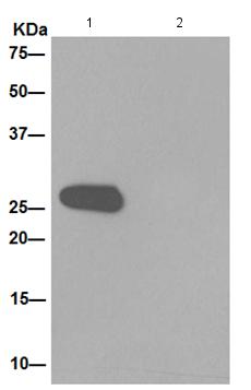
Western blot analysis on immunoprecipitation from 1) Jurkat cell lysate and 2) PBS, labeling PSMA4 using ab191403 at 1/200 dilution and HRP-conjugated anti-rabbit IgG preferentially detecting the non-reduced form of rabbit IgG at a 1/1500 dilution.