![All lanes : Anti-PSF1 antibody [EPR13359] (ab181112) at 1/20000 dilutionLane 1 : Jurkat cell lysateLane 2 : HeLa cell lysateLane 3 : HepG2 cell lysateLysates/proteins at 20 µg per lane.SecondaryGoat Anti-Rabbit IgG, (H+L), Peroxidase conjugate at 1/1000 dilution](http://www.bioprodhub.com/system/product_images/ab_products/2/sub_4/17700_ab181112-215036-ab181112WB.jpg)
All lanes : Anti-PSF1 antibody [EPR13359] (ab181112) at 1/20000 dilutionLane 1 : Jurkat cell lysateLane 2 : HeLa cell lysateLane 3 : HepG2 cell lysateLysates/proteins at 20 µg per lane.SecondaryGoat Anti-Rabbit IgG, (H+L), Peroxidase conjugate at 1/1000 dilution
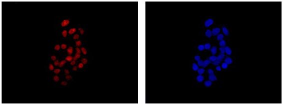
Immunofluorescent analysis of 4% paraformaldehyde-fixed HepG2 cells labeling PSF1 with ab181112 at 1/500 dilution followed by Goat anti rabbit IgG (Alexa Fluor® 555) secondary antibody at 1/200 dilution. Counter stained with Dapi (blue).

Western blot analysis of PSF1 in HepG2 cell lysate immunoprecipitated with ab181112 at 1/50 dilution (Lane 1). Lane 2: Negative control.Secondary: Anti-Rabbit IgG (HRP), specific to the non-reduced form of IgG at 1/1500 dilution.