![All lanes : Anti-PRPF4 antibody [EPR17206(B)] (ab201684) at 1/5000 dilutionLane 1 : HeLa (Human epithelial cells from cervix adenocarcinoma) whole cell lysateLane 2 : SW480 (Human colon adenocarcinoma cell line) whole cell lysateLane 3 : Raji (Human Burkitt's lymphoma cell line) whole cell lysateLane 4 : Jurkat (Human T cell leukemia cells from peripheral blood) whole cell lysateLysates/proteins at 20 µg per lane.SecondaryGoat Anti-Rabbit IgG, (H+L), Peroxidase conjugated at 1/1000 dilution](http://www.bioprodhub.com/system/product_images/ab_products/2/sub_4/17319_ab201684-244333-ab201684-wb-1.jpg)
All lanes : Anti-PRPF4 antibody [EPR17206(B)] (ab201684) at 1/5000 dilutionLane 1 : HeLa (Human epithelial cells from cervix adenocarcinoma) whole cell lysateLane 2 : SW480 (Human colon adenocarcinoma cell line) whole cell lysateLane 3 : Raji (Human Burkitt's lymphoma cell line) whole cell lysateLane 4 : Jurkat (Human T cell leukemia cells from peripheral blood) whole cell lysateLysates/proteins at 20 µg per lane.SecondaryGoat Anti-Rabbit IgG, (H+L), Peroxidase conjugated at 1/1000 dilution
![Anti-PRPF4 antibody [EPR17206(B)] (ab201684) at 1/5000 dilution + Human fetal spleen lysate at 10 µgSecondaryAnti-Rabbit IgG (HRP), specific to the non-reduced form of IgG at 1/1000 dilution](http://www.bioprodhub.com/system/product_images/ab_products/2/sub_4/17320_ab201684-244332-ab201684-wb-2.jpg)
Anti-PRPF4 antibody [EPR17206(B)] (ab201684) at 1/5000 dilution + Human fetal spleen lysate at 10 µgSecondaryAnti-Rabbit IgG (HRP), specific to the non-reduced form of IgG at 1/1000 dilution
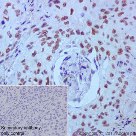
Immunohistochemical analysis of paraffin-embedded Human cervix carcinoma tissue labeling PRPF4 with ab201684 at 1/3000 dilution, followed by Goat Anti-Rabbit IgG H&L (HRP) (ab97051) secondary antibody at 1/500 dilution. Nuclear staining on Human cervix carcinoma tissue is observed. Counter stained with Hematoxylin.Secondary antibody only control: Used PBS instead of primary antibody, secondary antibody is Goat Anti-Rabbit IgG H&L (HRP) (ab97051) at 1/500 dilution.
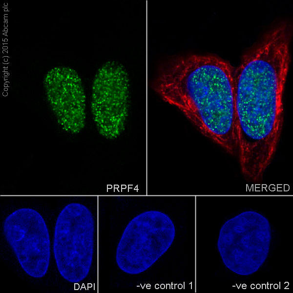
Immunofluorescent analysis of 4% paraformaldehyde-fixed, 0.1% Triton X-100 permeabilized HeLa (Human epithelial cells from cervix adenocarcinoma) cells labeling PRPF4 with ab201684 at 1/1200 dilution, followed by Goat anti-rabbit IgG (Alexa Fluor® 488) (ab150077) secondary antibody at 1/500 dilution (green). Confocal image showing nuclear staining on HeLa cell line. The nuclear counter stain is DAPI (blue). Tubulin is detected with ab7291 (anti-Tubulin mouse mAb) at 1/1000 dilution and ab150120 (AlexaFluor®594 Goat anti-Mouse secondary) at 1/500 dilution (red).The negative controls are as follows:-ve control 1: ab201684 at 1/1200 dilution followed by ab150120 (AlexaFluor®594 Goat anti-Mouse secondary) at 1/500 dilution.-ve control 2: ab7291 (anti-Tubulin mouse mAb) at 1/1000 dilution followed by ab150077 (Alexa Fluor®488 Goat Anti-Rabbit IgG H&L) at 1/500 dilution.
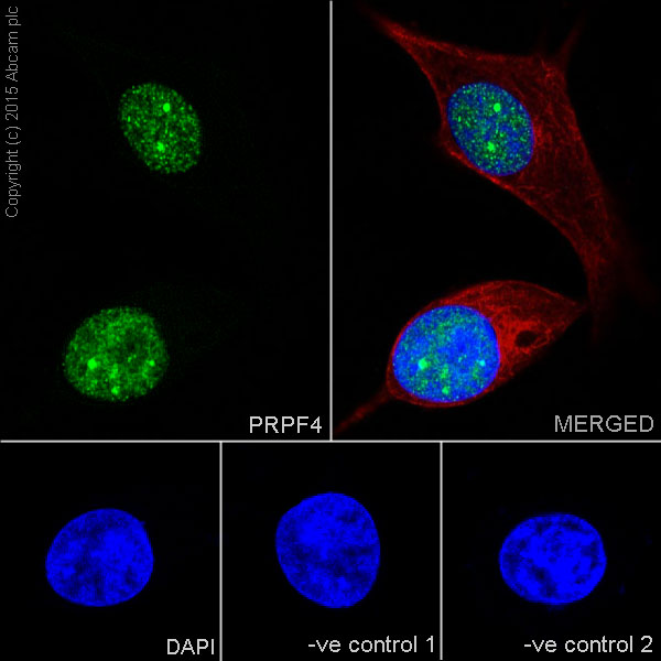
Immunofluorescent analysis of 4% paraformaldehyde-fixed, 0.1% Triton X-100 permeabilized MCF7 (Human breast adenocarcinoma cell line) cells labeling PRPF4 with ab201684 at 1/1200 dilution, followed by Goat anti-rabbit IgG (Alexa Fluor® 488) (ab150077) secondary antibody at 1/500 dilution (green). Confocal image showing nuclear staining on MCF7 cell line. The nuclear counter stain is DAPI (blue). Tubulin is detected with ab7291 (anti-Tubulin mouse mAb) at 1/1000 dilution and ab150120 (AlexaFluor®594 Goat anti-Mouse secondary) at 1/500 dilution (red).The negative controls are as follows:-ve control 1: ab201684 at 1/1200 dilution followed by ab150120 (AlexaFluor®594 Goat anti-Mouse secondary) at 1/500 dilution.-ve control 2: ab7291 (anti-Tubulin mouse mAb) at 1/1000 dilution followed by ab150077 (Alexa Fluor®488 Goat Anti-Rabbit IgG H&L) at 1/500 dilution.
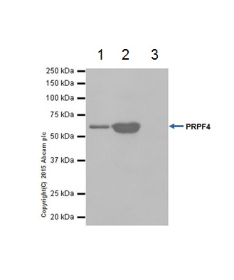
PRPF4 was immunoprecipitated from 1mg of HeLa (Human epithelial cells from cervix adenocarcinoma) whole cell lysate with ab201684 at 1/40 dilution. Western blot was performed from the immunoprecipitate using ab201684 at 1/1000 dilution. VeriBlot for IP secondary antibody (HRP) (ab131366) was used as secondary antibody at 1/1500 dilution.Lane 1: HeLa whole cell lysate 10 µg (Input). Lane 2: ab201684 IP in HeLa whole cell lysate. Lane 3: Rabbit monoclonal IgG (ab172730) instead of ab201684 in HeLa whole cell lysate.Blocking and dilution buffer and concentration: 5% NFDM/TBST.Exposure time: 5 seconds.