Anti-Presenilin 2 antibody [APS 26]
| Name | Anti-Presenilin 2 antibody [APS 26] |
|---|---|
| Supplier | Abcam |
| Catalog | ab15549 |
| Prices | $387.00 |
| Sizes | 200 µg |
| Host | Mouse |
| Clonality | Monoclonal |
| Isotype | IgG1 |
| Clone | APS 26 |
| Applications | ICC/IF ICC/IF IP ICC/IF IHC-P WB ELISA |
| Species Reactivities | Mouse, Rat, Human, Bovine, Pig, Primate |
| Antigen | Synthetic peptide corresponding to Human Presenilin 2 aa 317-334 |
| Description | Mouse Monoclonal |
| Gene | PSEN2 |
| Conjugate | Unconjugated |
| Supplier Page | Shop |
Product images
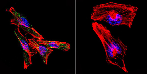
 Immunocytochemistry/Immunofluorescence analysis of HeLa cells labeling Presenilin 2 (green) with ab15549 at 1/20. F-Actin staining with Phalloidin (red) and nuclei with DAPI (blue). Cells were fixed with formaldehyde and incubated with the primary antibody overnight at 4°C. A DyLight 488-conjugated secondary antibody was used. 60X magnification. Right - negative control.
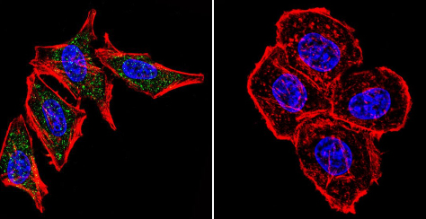
 Immunocytochemistry/Immunofluorescence analysis of A2058 cells labeling Presenilin 2 (green) with ab15549 at 1/20. F-Actin staining with Phalloidin (red) and nuclei with DAPI (blue). Cells were fixed with formaldehyde and incubated with the primary antibody overnight at 4°C. A DyLight 488-conjugated secondary antibody was used. 60X magnification. Right - negative control.
 IF staining Presenilin 2 in Mouse fibroblasts using ab15549
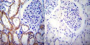 Immunohistochemistry was performed on normal biopsies of deparaffinized Human kidney tissue. To expose target proteins heat induced antigen retrieval was performed using 10mM sodium citrate (pH6.0) buffer microwaved for 8-15 minutes. Following antigen retrieval tissues were blocked in 3% BSA-PBS for 30 minutes at room temperature. Tissues were then probed at a dilution of 1:20 with a mouse monoclonal antibody recognizing Presenilin 2 ab15549 or without primary antibody (negative control) overnight at 4°C in a humidified chamber. Tissues were washed extensively with PBST and endogenous peroxidase activity was quenched with a peroxidase suppressor. Detection was performed using a biotin-conjugated secondary antibody and SA-HRP followed by colorimetric detection using DAB. Tissues were counterstained with hematoxylin and prepped for mounting.
Product References
Thapsigargin affects presenilin-2 but not presenilin-1 regulation in SK-N-BE - Thapsigargin affects presenilin-2 but not presenilin-1 regulation in SK-N-BE
Rivabene R, Visentin S, Piscopo P, De Nuccio C, Crestini A, Svetoni F, Rosa P, Confaloni A. Exp Biol Med (Maywood). 2014 Feb;239(2):213-24.
Interactome mapping suggests new mechanistic details underlying Alzheimer's - Interactome mapping suggests new mechanistic details underlying Alzheimer's
Soler-Lopez M, Zanzoni A, Lluis R, Stelzl U, Aloy P. Genome Res. 2011 Mar;21(3):364-76.