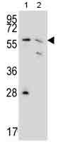
All lanes : Anti-PGD antibody (ab175116) at 1/1000 dilutionLane 1 : 293 cell lysateLane 2 : MCF7 cell lysateLysates/proteins at 35 µg per lane.

Anti-PGD antibody (ab175116) at 1/1000 dilution + Mouse spleen tissue lysate at 35 µg

Flow cytometric analysis of 293 cells (right histogram) compared to a negative control cell (left histogram) labeling PGD with ab175116 at 1/10 dilution. FITC-conjugated goat-anti-rabbit secondary antibodies were used for the analysis.

Immunohistochemistry analysis of formalin fixed and paraffin embedded Human lung carcinoma tissue labeling PGD with ab175116 at 1/50 dilution, followed by peroxidase conjugation of the secondary antibody and DAB staining.