
Anti-PFKFB4 antibody (ab71622) at 1/100 dilution + mouse brain tissue lysate at 12.5 µg

Formalin-fixed and paraffin-embedded human hepatocarcinoma tissue reacted with ab71622 at a 1:50 dilution. A peroxidase-conjugated secondary antibody was then used, followed by AEC staining.

Formalin-fixed and paraffin-embedded human hepatocarcinoma tissue reacted with ab71622 at 1:50 dilution. A peroxidase-conjugated secondary antibody was then used, followed by DAB staining.
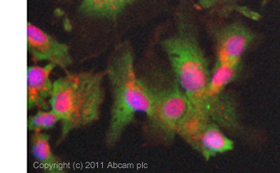
ICC/IF image of ab71622 stained HepG2 cells. The cells were 4% PFA fixed (10mins) and then incubated in 1%BSA / 10% normal goat serum / 0.3M glycine in 0.1% PBS-Tween for 1h to permeabilise the cells and block non-specific protein-protein interactions. The cells were then incubated with the antibody (ab71622, 5µg/ml) overnight at +4°C. The secondary antibody (green) was ab96899 Dylight 488 goat anti-rabbit IgG (H+L) used at a 1/250 dilution for 1h. Alexa Fluor® 594 WGA was used to label plasma membranes (red) at a 1/200 dilution for 1h. DAPI was used to stain the cell nuclei (blue) at a concentration of 1.43µM.