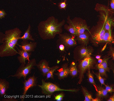
ICC/IF image of ab70175 stained HepG2 cells. The cells were 100% methanol fixed (5 min) and then incubated in 1%BSA / 10% normal goat serum / 0.3M glycine in 0.1% PBS-Tween for 1h to permeabilise the cells and block non-specific protein-protein interactions. The cells were then incubated with the antibody ab70175 at 5µg/ml overnight at +4°C. The secondary antibody (green) was DyLight® 488 goat anti- rabbit (ab96899) IgG (H+L) used at a 1/250 dilution for 1h. Alexa Fluor® 594 WGA was used to label plasma membranes (red) at a 1/200 dilution for 1h. DAPI was used to stain the cell nuclei (blue) at a concentration of 1.43µM.
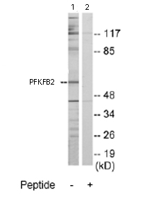
All lanes : Anti-PFKFB2 antibody (ab70175) at 1/500 dilutionLane 1 : Extracts from Jurkat cellsLane 2 : Extracts from Jurkat cells with immunising peptide at 10 µgLysates/proteins at 30 µg per lane.
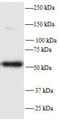
Anti-PFKFB2 antibody (ab70175) at 1/500 dilution (in 5% Marvel TBS-T for 24 hours at 22°C) + Whole cell lysate of mouse NIH 3T£ cells at 25 µgSecondaryAn HRP-conjugated Sheep anti-rabbit monoclonal at 1/5000 dilution