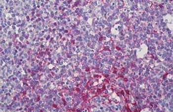
Immunohistochemical analysis of formalin-fixed, paraffin-embedded Human tonsil tissue labelling PDLIM1 with ab133711 at 15 µg/ml, followed by biotinylated goat anti-rabbit IgG secondary antibody, alkaline phosphatase-streptavidin and chromogen.
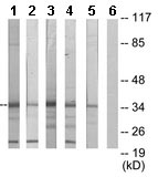
All lanes : Anti-PDLIM1 antibody (ab133711) at 1/500 dilutionLane 1 : Jurkat cell extractLane 2 : COLO205 cell extractLane 3 : 293 cell extractLane 4 : HepG2 cell extractLane 5 : HuvEc cell extractLane 6 : HuvEc cell extract with immunizing peptide
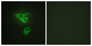
Immunofluorescent analysis of HepG2 cells labelling PDLIM1 with ab133711 at 1/100 dilution. Right panel is treated with the immunizing peptide.