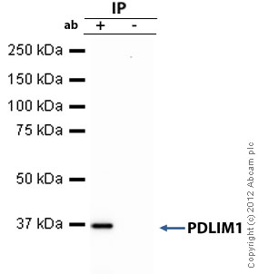
PDLIM1 was immunoprecipitated using 0.5mg HepG2 whole cell extract, 5µg of Rabbit polyclonal to PDLIM1 and 50µl of protein G magnetic beads (+). No antibody was added to the control (-). The antibody was incubated under agitation with Protein G beads for 10min, HepG2 whole cell extract lysate diluted in RIPA buffer was added to each sample and incubated for a further 10min under agitation.Proteins were eluted by addition of 40µl SDS loading buffer and incubated for 10min at 70oC; 10µl of each sample was separated on a SDS PAGE gel, transferred to a nitrocellulose membrane, blocked with 5% BSA and probed with ab64971.Secondary: Goat polyclonal to mouse IgG light chain specific (HRP) at 1/5000 dilution.Band: 36kDa: PDLIM1.
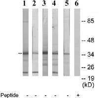
All lanes : Anti-PDLIM1 antibody (ab64971) at 1/500 dilutionLane 1 : Jurkat cell extract; without immunizing peptideLane 2 : COLO205 cell extract; without immunizing peptideLane 3 : 293 cell extract; without immunizing peptideLane 4 : HepG2 cell extract; without immunizing peptideLane 5 : HUVEC cell extract; without immunizing peptideLane 6 : HUVEC cell extract; with immunizing peptide
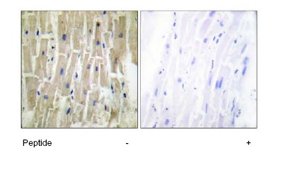
Immunohistochemical analysis of PDLIM1 in in paraffin embedded human heart tissue using ab64971 at 1/50 dilution; in the presence (left) and absence (right) of the immunizing peptide.
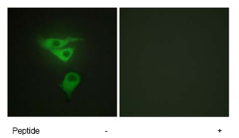
Immunofluorescence analysis of PDLIM1 in HepG2 cells, with ab64971 (1/500 diltion). in the presence (left) and absence (right) of the immunizing peptide.
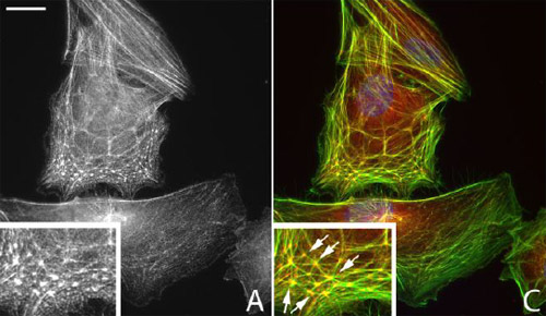
Immunofluorescence analysis of Human trabecular meshwork cells, staining PDLIM1 with ab64971. Left panel: PDLIM1 staining alone. Right panel: PDLIM1 staining (red) merged with F-actin staining (green). Cells were treated with dexamethasone, before fixing in paraformaldehyde and permeabilizing wih 0.2% Triton X-100. Cells were incubated with primary antibody (2 µg/ml) and AlexaFluor®488-conjugated phalloidin. PDLIM1 staining was detected using an AlexaFluor®546-conjugated goat anti-rabbit IgG.